. 2010 Jan;38(3):1009-21.
doi: 10.1093/nar/gkp1029. Epub 2009 Nov 27.

Structure of a two-G-tetrad intramolecular G-quadruplex formed by a variant human telomeric sequence in K+ solution: insights into the interconversion of human telomeric G-quadruplex structures

Affiliations

Structure of a two-G-tetrad intramolecular G-quadruplex formed by a variant human telomeric sequence in K+ solution: insights into the interconversion of human telomeric G-quadruplex structures

Zhenjiang Zhang et al. Nucleic Acids Res. 2010 Jan.

Abstract

Human telomeric DNA G-quadruplex has been considered as an attractive target for cancer therapeutic intervention. The telomeric sequence shows intrinsic structure polymorphism. Here we report a novel intramolecular G-quadruplex structure formed by a variant human telomeric sequence in K(+) solution. This sequence forms a basket-type intramolecular G-quadruplex with only two G-tetrads but multiple-layer capping structures formed by loop residues. While it is shown that this structure can only be detected in the specifically truncated telomeric sequences without any 5'-flanking residues, our results suggest that this two-G-tetrad conformation is likely to be an intermediate form of the interconversion of different telomeric G-quadruplex conformations.


Figures
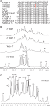
Figure 1.
Figure 1.
(A) Native and variant four-G-tract human telomeric DNA sequences. Tel21 is used as the base sequence to show various flanking segments. The numbering system is shown above A–Tel21–T. The sequences used for structure determination are labeled, with the structure determined in parentheses. (B) Imino regions of 1D 1H NMR spectra of several telomeric sequences shown in (A). The assignment for I14–Tel23 is shown. The G10NH1 proton of Tel21–T and A–Tel21–T are labeled with asterisks. (C) The aromatic region of 1D 1H NMR spectra of I14–Tel23 in K+ solutions at 20°C, with assignments. Conditions: 25 mM KPO4, 70 mM KCl, pH 7.0, 20°C.
Figure 2.
Figure 2.
The imino proton region with assignment of the 1D 1H NMR spectrum of I14–Tel23 in K+ solution (top), and imino proton assignments of I14–Tel23 using 1D 15N-filtered experiments on site-specific labeled oligonucleotides. Conditions: 25 mM KPO4, 70 mM KCl, pH 7.0, 20°C, 0.50–0.6 mM DNA. G4NH1 was assigned using the I14–Tel22 sequence.
Figure 3.
Figure 3.
(A) The expanded H1–H8/H2/H6 (top) and H1–H1 (bottom) regions, with labeling, of the exchangeable proton 2D JR-NOESY spectrum of I14–Tel23 in K+ solution at 1°C. Only the aromatic H2 protons are specified in the labels. NOEs are labeled as following: intra-tetrad NOEs are labeled in red, inter-tetrad NOEs are labeled in purple, intra-triple NOEs are labeled in green, NOEs between the G-tetrads and the stacking triple structures are labeled in blue. (B) The expanded H8/H6–H1′ region of the non-exchangeable 2D-NOESY spectrum of I14–Tel23 in K+ solution at 20°C. The sequential assignment pathway is shown. The H8–H1′ NOEs are labeled with residue names. The residue names of the guanines with syn conformation are in red. A3 and A15 show much broader peaks, likely related with the dynamic motion. Missing connectivities are labeled with arrows. The characteristic G(i)H8/G(i + 1)H1′ NOEs for the syn G(i)s are labeled in green. Conditions: 25 mM pH 7.0 K-phosphate, 70 mM KCl, 2.0 mM DNA.
Figure 4.
Figure 4.
(A) Schematic drawing of the folding topologies of the intramolecular G-quadruplex formed in I14–Tel23 in K+ solution (right). I14 is in orange. Red box = (anti) guanine, purple box = (syn) guanine, green box = adenine, blue box = thymine; red ball = guanine, green ball = adenine, blue ball = thymine. A G-tetrad with H1–H1 and H1–H8 connectivity patterns detectable in NOESY experiments is also shown in the inset. (B) Schematic drawing of the G10–I14–G22 and G4–A7–A19 triple structures with potential H-bonds. (C) CD spectra of several telomeric sequences shown in Figure 1A in pH7 K+ or Na+ solution at 25°C.
Figure 5.
Figure 5.
Schematic diagram of inter-residue NOE connectivities of I14–Tel23 G-quadruplex formed in K+ solution. The guanines in syn conformation are represented using gray circles. The NOE connectivities clearly define the G-quadruplex conformation and provide distance restraints for structure calculation.
Figure 6.
Figure 6.
(A) Stereo view of the superimposed 8 NMR-refined structures of the two-G-tetrad G-quadruplex formed in I14–Tel23 in K+ solution. The G2–G15–G21–G9 tetrad is in light pink, and the G3–G16–G20–G8 is in yellow. The guanines in the G10–I14–G22 and G4–A7–A19 triples are in magenta, I14 in orange, adenines are in red, and thymines in blue. (B) A representative model of the NMR-refined I14–Tel23 G-quadruplex structure from two different views, prepared using GRASP (43) (guanine = yellow, adenine = red, thymine = blue).
Figure 7.
Figure 7.
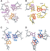
Loop conformations in the two-G-tetrad G-quadruplex formed by I14–Tel23 in K+ solution. (A) The top view (as in Figure 6) of the G10–I14–G22 triple capping the G2–G15–G21–G9 G-tetrad. (B) The bottom view of the G4–A7–A19 triple capping the G3–G16–G20–G8 G-tetrad. (C) The top view of the T12–T23 base pair capping the G10–I14–G22 triple. (D) The bottom view of the T6–T18 base pair capping the G4–A7–A19 triple. The color code is the same as that in Figure 6A.
Figure 8.
Figure 8.
Schematic diagram of a proposed model for interconversions between the Na+ basket form (A) and the K+ hybrid form of telomeric G-quadruplexes (D and D2), and between the two hybrid forms telomeric G-quadruplexes (D and D2). Guanines with known anti conformation are in red, and guanines with syn conformation are in purple. The A–Tel21 forms a single stable basket-type intramolecular G-quadruplex in Na+ solution. In the presence of K+ the basket form (A) is not favored and A–Tel21 can convert to the hybrid forms (D and D2) through a strand-reorientation mechanism (C and C2). The two-G-tetrad conformation is likely to be the intermediate state of this strand reorientation and may be stabilized by specific loop interactions of a specific sequence. For example, the presence of I14 loosens the H-bonding connection between G14 and G2 in the basket-form (A) and thus frees the 5′-end for its sliding-up dissociation from the core G-tetrads. However, this strand sliding-up reorientation is captured at the two-G-tetrad stage (B) due to the stable formation of the G10–I14–G22 capping structure. A similar sliding (B2) and dissociation (C2) could also potentially happen at the 3′-end of the basket-form structure to convert to the hybrid-2 form G-quadruplex by the 3′-end swing-back (D2). The two-G-tetrad form may also be an intermediate state of the interconversion between the two hybrid-type forms (D↔C↔B↔B2↔C2↔D2 circle).

Similar articles

Cited by

References

    1. Blackburn EH. Telomere states and cell fates. Nature. 2000;408:53–56. - PubMed
    1. van Steensel B, Smogorzewska A, de Lange T. TRF2 protects human telomeres from end-to-end fusions. Cell. 1998;92:401–413. - PubMed
    1. Hackett JA, Feldser DM, Greider CW. Telomere dysfunction increases mutation rate and genomic instability. Cell. 2001;106:275–286. - PubMed
    1. Mergny JL, Helene C. G-quadruplex DNA: A target for drug design. Nat. Med. 1998;4:1366–1367. - PubMed
    1. Sun DY, Hurley LH. Targeting telomeres and telomerase. In: Chaires JB, Waring MJ, editors. Methods in Enzymology, Drug-Nucleic Acid Interactions. Vol. 340. San Diego: Academic Press Inc; 2001. pp. 573–592. - PubMed

Publication types