

Mechanism of Wound-Healing Activity of Hippophae rhamnoides L. Leaf Extract in Experimental Burns
- PMID: 19946025
- PMCID: PMC3152935
- DOI: 10.1093/ecam/nep189
Mechanism of Wound-Healing Activity of Hippophae rhamnoides L. Leaf Extract in Experimental Burns
Abstract
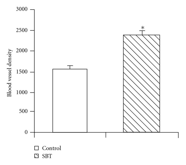
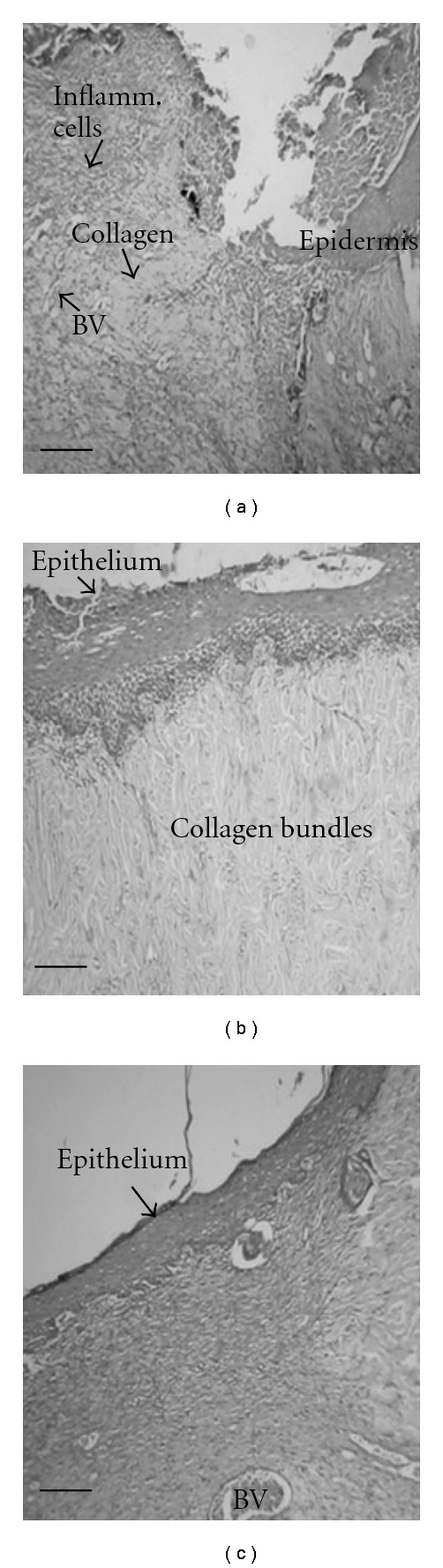
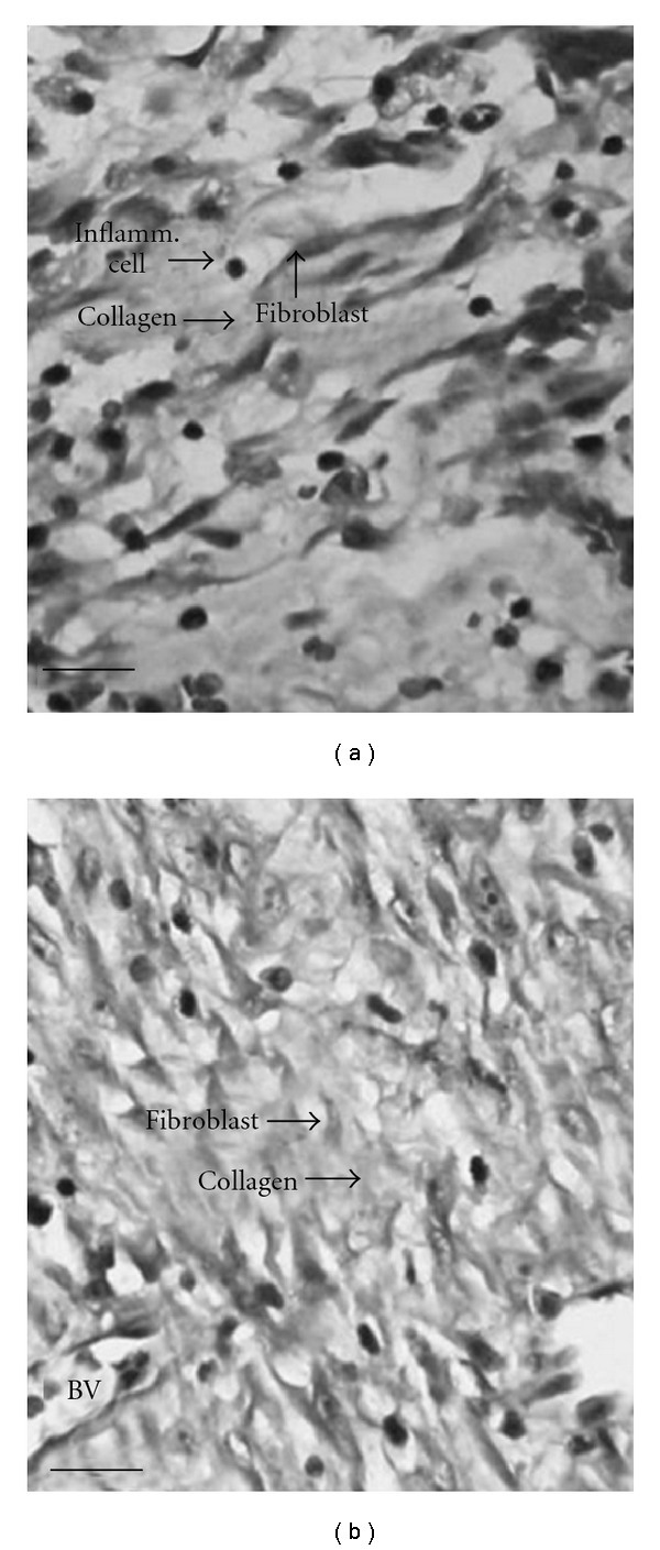
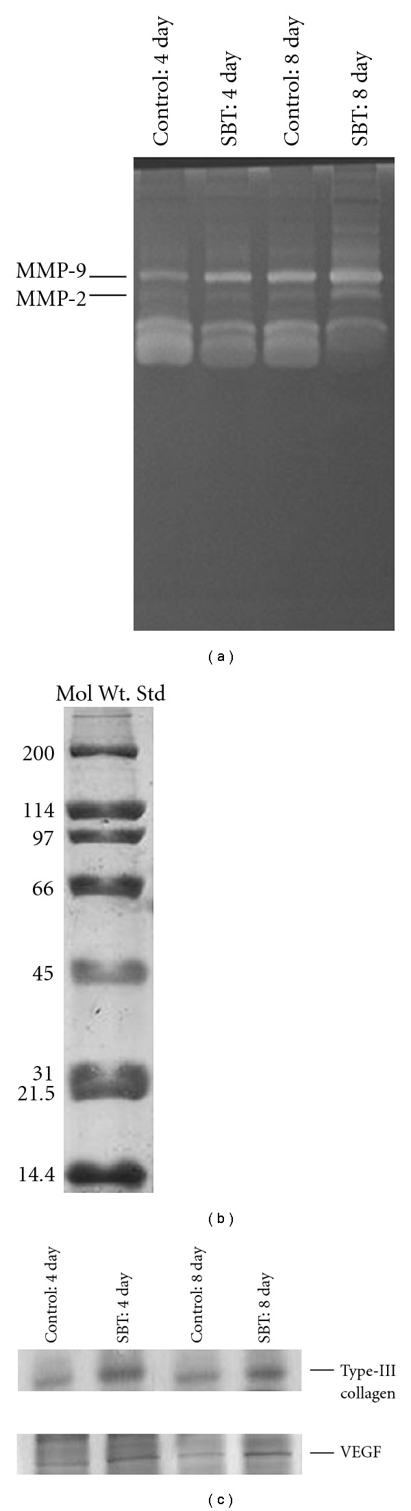
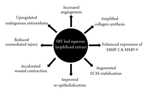
The present investigation was undertaken to evaluate the healing efficacy of lyophilized aqueous leaf extract of Sea buckthorn (Hippophae rhamnoides L., family Elaeagnaceae) (SBT) and to explore its possible mechanism of action on experimental burn wounds in rats. The SBT extract, at various concentrations, was applied topically, twice daily for 7 days. Treatment with silver sulfadiazine (SSD) ointment was used as reference control. The most effective concentration of the extract was found to be 5.0% (w/w) for burn wound healing and this was further used for detailed study. The SBT-treated group showed faster reduction in wound area in comparison with control and SSD-treated groups. The topical application of SBT increased collagen synthesis and stabilization at the wound site, as evidenced by increase in hydroxyproline, hexosamine levels and up-regulated expression of collagen type-III. The histological examinations and matrix metalloproteinases (MMP-2 and -9) expression also confirmed the healing efficacy of SBT leaf extract. Furthermore, there was significant increase in levels of endogenous enzymatic and non-enzymatic antioxidants and decrease in lipid peroxide levels in SBT-treated burn wound granulation tissue. The SBT also promoted angiogenesis as evidenced by an in vitro chick chorioallantoic membrane model and in vivo up-regulated vascular endothelial growth factor (VEGF) expression. The SBT leaf extract had no cytotoxic effect on BHK-21 cell line. In conclusion, SBT aqueous leaf extract possesses significant healing potential in burn wounds and has a positive influence on the different phases of wound repair.
Figures
Similar articles
-
Safety and healing efficacy of Sea buckthorn (Hippophae rhamnoides L.) seed oil on burn wounds in rats.Food Chem Toxicol. 2009 Jun;47(6):1146-53. doi: 10.1016/j.fct.2009.02.002. Food Chem Toxicol. 2009. PMID: 19425187
-
A preclinical study of the effects of seabuckthorn (Hippophae rhamnoides L.) leaf extract on cutaneous wound healing in albino rats.Int J Low Extrem Wounds. 2005 Jun;4(2):88-92. doi: 10.1177/1534734605277401. Int J Low Extrem Wounds. 2005. PMID: 15911921
-
Hippophae rhamnoides L. leaf extract augments dermal wound healing in streptozocin-induced diabetic rats.J Wound Care. 2025 Feb 2;34(2):146-153. doi: 10.12968/jowc.2021.0309. J Wound Care. 2025. PMID: 39928468
-
Influence of sea buckthorn (Hippophae rhamnoides L.) flavone on dermal wound healing in rats.Mol Cell Biochem. 2006 Oct;290(1-2):193-8. doi: 10.1007/s11010-006-9187-6. Epub 2006 Apr 22. Mol Cell Biochem. 2006. PMID: 16633732
-
Medicinal plants and their natural components as future drugs for the treatment of burn wounds: an integrative review.Arch Dermatol Res. 2014 Sep;306(7):601-17. doi: 10.1007/s00403-014-1474-6. Epub 2014 Jun 4. Arch Dermatol Res. 2014. PMID: 24895176 Review.
Cited by
-
Discovery of Anti-Coronavirus Cinnamoyl Triterpenoids Isolated from Hippophae rhamnoides during a Screening of Halophytes from the North Sea and Channel Coasts in Northern France.Int J Mol Sci. 2023 Nov 22;24(23):16617. doi: 10.3390/ijms242316617. Int J Mol Sci. 2023. PMID: 38068938 Free PMC article.
-
The Therapeutic Application of Tamarix aphylla Extract Loaded Nanoemulsion Cream for Acid-Burn Wound Healing and Skin Regeneration.Medicina (Kaunas). 2022 Dec 23;59(1):34. doi: 10.3390/medicina59010034. Medicina (Kaunas). 2022. PMID: 36676658 Free PMC article.
-
Rapid and selective mobilization of specific stem cell types after consumption of a polyphenol-rich extract from sea buckthorn berries (Hippophae) in healthy human subjects.Clin Interv Aging. 2019 Feb 4;14:253-263. doi: 10.2147/CIA.S186893. eCollection 2019. Clin Interv Aging. 2019. PMID: 30787601 Free PMC article. Clinical Trial.
-
Effect of piracetam and nimodipine on full-thickness skin burns in rabbits.Int Wound J. 2016 Aug;13(4):563-71. doi: 10.1111/iwj.12478. Epub 2015 Jul 20. Int Wound J. 2016. PMID: 26192365 Free PMC article.
-
Botanical Drugs in Traditional Chinese Medicine With Wound Healing Properties.Front Pharmacol. 2022 May 12;13:885484. doi: 10.3389/fphar.2022.885484. eCollection 2022. Front Pharmacol. 2022. PMID: 35645789 Free PMC article. Review.
References
-
- Singer AJ, Clark RAF. Cutaneous wound healing. The New England Journal of Medicine. 1999;341(10):738–746. - PubMed
-
- Gupta A, Upadhyay NK, Sawhney RC, Kumar R. A poly-herbal formulation accelerates normal and impaired diabetic wound healing. Wound Repair and Regeneration. 2008;16(6):784–790. - PubMed
-
- Arturson G. Pathophysiology of the burn wound and pharmacological treatment. The Rudi Hermans Lecture, 1995. Burns. 1996;22(4):255–274. - PubMed
-
- Thomas GW, Rael LT, Bar-Or R, et al. Mechanisms of delayed wound healing by commonly used antiseptics. Journal of Trauma. 2009;66(1):82–90. - PubMed
-
- Lee A-RC, Leem H, Lee J, Park KC. Reversal of silver sulfadiazine-impaired wound healing by epidermal growth factor. Biomaterials. 2005;26(22):4670–4676. - PubMed
LinkOut - more resources
Full Text Sources
Miscellaneous
