. 2009 Dec;212(Pt 24):3961-76.
doi: 10.1242/jeb.035741.

The peptide hormone pQDLDHVFLRFamide (crustacean myosuppressin) modulates the Homarus americanus cardiac neuromuscular system at multiple sites

Affiliations

The peptide hormone pQDLDHVFLRFamide (crustacean myosuppressin) modulates the Homarus americanus cardiac neuromuscular system at multiple sites

J S Stevens et al. J Exp Biol. 2009 Dec.

Abstract

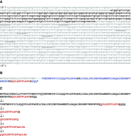
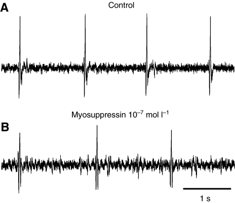
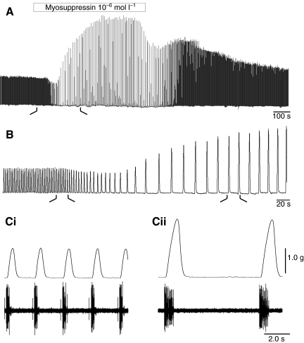
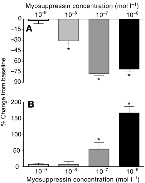
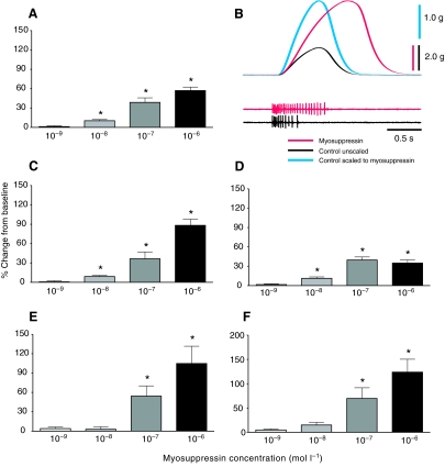
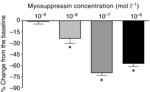
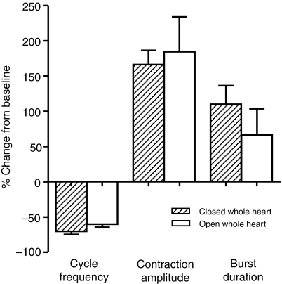
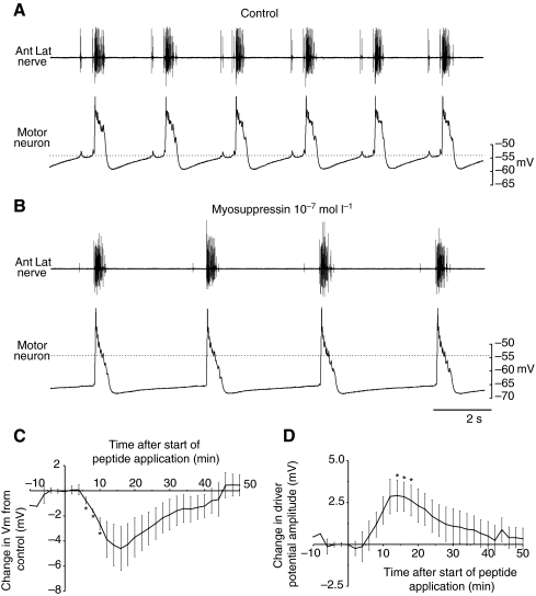
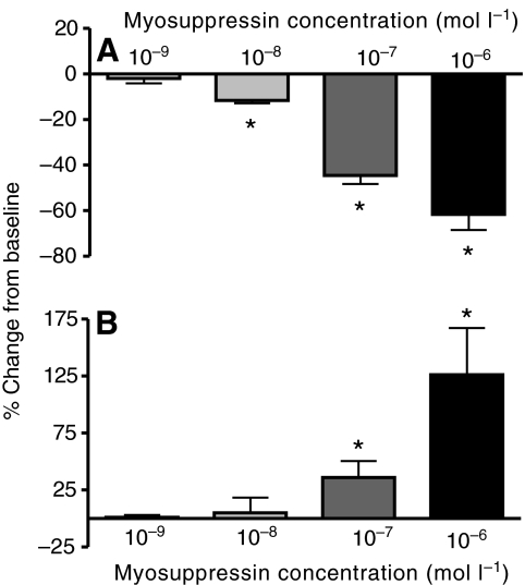
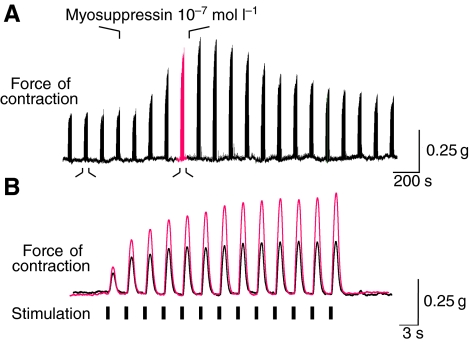
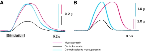
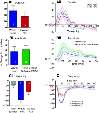
pQDLDHVFLRFamide is a highly conserved crustacean neuropeptide with a structure that places it within the myosuppressin subfamily of the FMRFamide-like peptides. Despite its apparent ubiquitous conservation in decapod crustaceans, the paracrine and/or endocrine roles played by pQDLDHVFLRFamide remain largely unknown. We have examined the actions of this peptide on the cardiac neuromuscular system of the American lobster Homarus americanus using four preparations: the intact animal, the heart in vitro, the isolated cardiac ganglion (CG), and a stimulated heart muscle preparation. In the intact animal, injection of myosuppressin caused a decrease in heartbeat frequency. Perfusion of the in vitro heart with pQDLDHVFLRFamide elicited a decrease in the frequency and an increase in the amplitude of heart contractions. In the isolated CG, myosuppressin induced a hyperpolarization of the resting membrane potential of cardiac motor neurons and a decrease in the cycle frequency of their bursting. In the stimulated heart muscle preparation, pQDLDHVFLRFamide increased the amplitude of the induced contractions, suggesting that myosuppressin modulates not only the CG, but also peripheral sites. For at least the in vitro heart and the isolated CG, the effects of myosuppressin were dose-dependent (10(-9) to 10(-6) mol l(-1) tested), with threshold concentrations (10(-8)-10(-7) mol l(-1)) consistent with the peptide serving as a circulating hormone. Although cycle frequency, a parameter directly determined by the CG, consistently decreased when pQDLDHVFLRFamide was applied to all preparation types, the magnitudes of this decrease differed, suggesting the possibility that, because myosuppressin modulates the CG and the periphery, it also alters peripheral feedback to the CG.


Figures

Fig. 1.
Fig. 1.
Nucleotide and deduced amino acid sequences of Homarus americanus prepro-myosuppressin. (A) Nucleotide sequence of Homarus americanus prepro-myosuppressin (accession no. GQ303179). The open reading frame of the cDNA, including the stop codon, is shown in bold font, with the 3′ polyadenylation signal sequences indicated by underline in black. (B) Deduced amino acid sequence of Homarus americanus prepro-myosuppressin. The signal peptide is shown in grey, with prohormone convertase cleavage loci shown in black. The encoded myosuppressin isoform is shown in red, with additional precursor-related peptides in blue. The asterisk indicates the position of the stop codon. (C) Putative processing scheme resulting in the production of myosuppressin from its precursor protein. The first line of sequence shows the full-length prepro-hormone with the locus of cleavage locus for signal peptidase underlined. The second line of sequence shows the cleaved pro-hormone with the loci for prohormone convertase underlined. The third line of sequence shows the immature myosuppressin peptide with the site of action for carboxypeptidase underlined. The fourth line of sequence shows the peptide resulting from carboxypeptidase activity with the Gly destined for a-amidation underlined. The fifth line of sequence shows the amidated peptide with the Gln destined for cyclization underlined. The final line of sequence shows the putative, mature myosuppressin after full post-translational processing.
Fig. 2.
Fig. 2.
Myosuppressin decreased heart rate when injected into intact lobsters. Heart rate was monitored with extracellular electrodes implanted over the heart. (A) Control heart rate was approximately 60 beats min−1. (B) Recording taken 6 min after injection of 500 μl of 5×10⁻³ mol l−1 myosuppressin, calculated to bring the initial concentration of peptide in the pericardial cavity to approximately 10−7 mol l−1. Heart rate slowed to ~40 beats min−1.
Fig. 3.
Fig. 3.
Myosuppressin decreased heart rate and had complex effects on amplitude when perfused through an isolated whole heart at 10−7 mol l−1. (A) Recording on a slow time base illustrates a typical response to myosuppressin: heart rate decreased, amplitude of contractions first decreased slightly (~10% in the example shown), then dramatically increased to over 200% of its original value. (B) Higher speed recording of initial response, taken from time demarcated in A by the black lines. (C) Higher speed recordings of heartbeat and associated neural activity, as recorded extracellularly on the anterior lateral nerve in control (Ci) and 4 min after the onset of myosuppressin perfusion (Cii), as demarcated in B. Cycle frequency decreased, contraction amplitude increased, and both burst and contraction duration increased in the presence of 10−7 mol l−1 myosuppressin.
Fig. 4.
Fig. 4.
Both heartbeat frequency and contraction amplitude in the whole heart preparation changed in a dose-dependent manner in the presence of myosuppressin. Pooled data from preparations exposed to concentrations of myosuppressin ranging from 10−6 to 10−9 mol l−1 showed that heart rate decreased (A) and contraction amplitude increased (B), with larger effects at higher concentrations. Threshold for cycle frequency, defined as the concentration needed to produce a change significantly different from zero, was between 10−8 and 10−9 mol l−1; threshold for changes in amplitude was between 10−7 and 10−8 mol l−1. *Significantly different from zero, one-sample, two-tailed t-test, P<0.05. N=6, 6, 11, 14 for 10−9, 10−8, 10−7 and 10−6 mol l−1 myosuppressin applications. Error bars represent standard errors.
Fig. 5.
Fig. 5.
Myosuppressin caused dose-dependent increases in contraction duration in whole heart preparations, with similar effects on a range of parameters related to duration. (A) Duration of contractions, measured at half-amplitude, increased with increasing myosuppressin concentration. (B) Recordings of a single contraction in control saline (black) and in myosuppressin (red) illustrate the difference in time course and amplitude of heart beats. The recording in control saline has been scaled to the amplitude of the contraction in myosuppressin (blue), showing that the biggest change is in the rate and duration of the rising phase of the contraction. (C) Rise time, measured as the time from the onset of contraction to the maximum contraction, increased with increasing myosuppressin concentration. (D) Fall time, measured as the time from the peak of contraction to the return to baseline tension, increased to a lesser extent. (E) Burst duration, measured on the anterolateral nerves, likewise increased with increasing myosuppressin concentration. A,C—E are all shown on the same vertical scale to facilitate comparisons between graphs. (F) Number of spikes per burst similarly increased with increasing myosuppressin concentration; spike frequency (not shown) thus remained approximately constant. Threshold for contraction duration, rise time and fall time was between 10−8 and 10−9 mol l−1; threshold for burst duration and number of spikes per burst was between 10−7 and 10−8 mol l−1. *Significantly different from zero, one-sample, two-tailed t-test, P<0.05. N=6, 6, 11, 14 for 10−9, 10−8, 10−7 and 10−6 mol l−1 myosuppressin, respectively. Error bars represent standard errors.
Fig. 6.
Fig. 6.
Duty cycle, calculated as the contraction duration per cycle period, decreased as myosuppressin concentration increased to 10−7 mol l−1, after which it remained relatively constant. Both contraction duration and cycle period increased, but the increase in cycle period was greater, leading to a decrease in duty cycle. Threshold was between 10−8 and 10−9 mol l−1; * Significantly different from zero, one-sample, two-tailed t-test, P<0.05. N=6, 6, 11, 14 for 10−9, 10−8, 10−7 and 10−6 mol l−1 myosuppressin, respectively. Error bars represent standard errors.
Fig. 7.
Fig. 7.
Eliminating the stretch caused by the build-up of perfusion fluid in the heart as heart rate slowed did not alter heart rate, contraction amplitude or the duration of bursts of action potentials recorded on the anterolateral nerves within the whole heart. Stretch was eliminated by cutting a slit in the ventral wall of the heart (open whole heart). There were no significant differences between any of the parameters measured in the open whole heart and the more intact preparation (closed whole heart), t-test, P>0.05, N=12. Error bars represent standard errors.
Fig. 8.
Fig. 8.
Myosuppressin superfused over the isolated cardiac ganglion (CG) decreased cycle frequency. (A) Recording of extracellular activity on the anterolateral nerve (Ant lat nerve) and a simultaneous intracellular recording from a motor neuron. Dotted line shows membrane potential just before the driver potential, at about −55 mV. (B) In 10−7 mol l−1 myosuppressin, cycle frequency decreased, and the motor neuron hyperpolarized, in this case by approx. 12 mV. It can also be seen that the duration of the depolarizing slow wave (driver potential) in the motor neuron increased in myosuppressin. Dotted line indicates the membrane potential in control saline for comparison. (C,D) Pooled data from five preparations, illustrating the time course of the changes in membrane potential (C) and driver potential amplitude (D) after the start of myosuppressin superfusion (at time=0). Myosuppressin was superfused for 8 min before the preparation was returned to control saline. Error bars represent standard errors.
Fig. 9.
Fig. 9.
Both cycle frequency and burst duration in the isolated CG changed in a dose-dependent manner in the presence of myosuppressin. Pooled data from preparations exposed to concentrations of myosuppressin ranging from 10−6 to 10−9 mol l−1 showed that cycle frequency decreased (A) and burst duration increased (B), with larger effects at higher concentrations. Threshold for cycle frequency was between 10−8 and 10−9 mol l−1; threshold for the increase in burst duration was between 10−7 and 10−8 mol l−1. *Significantly different from zero, one-sample, two-tailed t-test, P<0.05, N=3, 3, 10, 6 for 10−9, 10−8, 10−7 and 10−6 mol l−1, respectively. Error bars represent standard errors.
Fig. 10.
Fig. 10.
Myosuppressin perfused through the heart at 10−7 mol l−1 caused an increase in the amplitude of nerve-evoked contractions. (A) One of the anterolateral nerves was stimulated with 300 ms bursts of 60 Hz stimuli. Each burst caused a single contraction of the heart. Bursts were delivered in bouts of 13 bursts once every 2 min. (A) On a slow time base, each bout is compressed to form a single peak; the amplitude of these peaks more than doubled when 10−7 mol l−1 myosuppressin was perfused through the heart. (B) Faster time scale illustrates one bout of bursts in control (black, smaller contractions) and in myosuppressin (red, larger amplitude contractions). In this example, there was a substantial increase in facilitation in myosuppressin, but statistically, facilitation did not increase across multiple preparations.
Fig. 11.
Fig. 11.
Comparison of the effects of 10−7 mol l−1 myosuppressin on the time courses of single heart contractions evoked by controlled nerve stimulation (A) with those generated by spontaneous neuronal activity (B). Control contractions are shown in black; myosuppressin are in red; control contractions scaled to the amplitude of myosuppressin contractions are in blue. Myosuppressin increased the duration of contractions elicited by controlled stimulation, but to a much smaller extent than those generated by spontaneous activity of the CG. Only the falling phase of stimulation-evoked contractions was increased by myosuppressin; both the rising and falling phases of CG-evoked contractions were slowed by myosuppressin.
Fig. 12.
Fig. 12.
Comparisons of the extents and time courses of the effects of 10−7 mol l−1 myosuppressin on burst duration, contraction amplitude and heart beat frequency in the four preparation types. (A) Burst duration was increased to similar extents (Ai) and with similar time courses (Aii) in the whole heart and the isolated CG. (B) Contraction amplitude was increased to similar extents (Bi) and with similar time courses (Bii) in the whole heart and in the heart stimulated with controlled pulses (nerve-evoked muscle contraction). (C) The effects of myosuppressin on heartbeat frequency differed significantly among preparation types (ANOVA, P<0.05). Post-hoc t-tests indicated that the values for all three types of preparation, i.e. the intact animal, the whole heart and the isolated CG, differed from each other (P<0.05), with significantly larger decreases in the whole heart than in the isolated CG, and larger decreases in the isolated CG than in the intact animal. Comparison of time courses in Ai, Bii and Cii suggests that the changes in cycle frequency peaked sooner than did changes in contraction amplitude. Different letters over bars indicate values that were significantly different from one another. N values are indicated on each bar in Ai, Bi and Ci. Stippled shading in Aii, Bii and Cii indicates standard errors.

Similar articles

Cited by

References

    1. Alexandrowicz J. S. (1932). Innervation of the heart of the Crustacea. I. Decapoda. Q. J. Microsc. Sci. 75, 182-249
    1. Altschul S. F., Madden T. L., Schäffer A. A., Zhang J., Zhang Z., Miller W., Lipman D. J. (1997). Gapped BLAST and PSI-BLAST: a new generation of protein database search programs. Nucleic Acids Res. 25, 3389-3402 - PMC - PubMed
    1. Anderson M., Cooke I. M. (1971). Neural activation of the heart of the lobster Homarus americanus. J. Exp. Biol. 55, 449-468 - PubMed
    1. Battelle B. A., Kravitz E. A. (1978). Targets of octopamine action in the lobster: cyclic nucleotide changes and physiological effects in hemolymph, heart and exoskeletal muscle. J. Pharmacol. Exp. Ther. 205, 438-448 - PubMed
    1. Bendtsen J. D., Nielsen H., von Heijne G., Brunak S. (2004). Improved prediction of signal peptides: SignalP 3. 0. J. Mol. Biol. 340, 783-795 - PubMed

Publication types

MeSH terms

Associated data