Review
. 2009 Dec;73(4):577-93.
doi: 10.1128/MMBR.00020-09.

ABC transporters in Saccharomyces cerevisiae and their interactors: new technology advances the biology of the ABCC (MRP) subfamily

Affiliations
Review

ABC transporters in Saccharomyces cerevisiae and their interactors: new technology advances the biology of the ABCC (MRP) subfamily

Christian M Paumi et al. Microbiol Mol Biol Rev. 2009 Dec.

Abstract

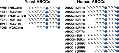
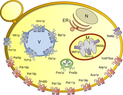
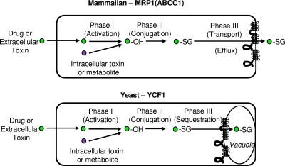
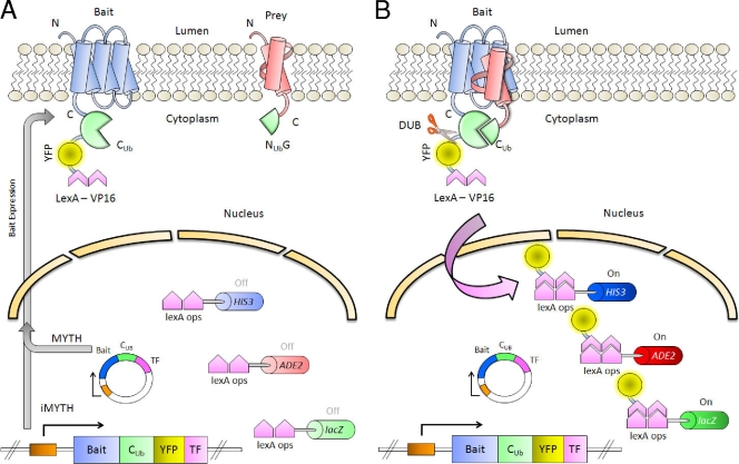
Members of the ATP-binding cassette (ABC) transporter superfamily exist in bacteria, fungi, plants, and animals and play key roles in the efflux of xenobiotic compounds, physiological substrates, and toxic intracellular metabolites. Based on sequence relatedness, mammalian ABC proteins have been divided into seven subfamilies, ABC subfamily A (ABCA) to ABCG. This review focuses on recent advances in our understanding of ABC transporters in the model organism Saccharomyces cerevisiae. We propose a revised unified nomenclature for the six yeast ABC subfamilies to reflect the current mammalian designations ABCA to ABCG. In addition, we specifically review the well-studied yeast ABCC subfamily (formerly designated the MRP/CFTR subfamily), which includes six members (Ycf1p, Bpt1p, Ybt1p/Bat1p, Nft1p, Vmr1p, and Yor1p). We focus on Ycf1p, the best-characterized yeast ABCC transporter. Ycf1p is located in the vacuolar membrane in yeast and functions in a manner analogous to that of the human multidrug resistance-related protein (MRP1, also called ABCC1), mediating the transport of glutathione-conjugated toxic compounds. We review what is known about Ycf1p substrates, trafficking, processing, posttranslational modifications, regulation, and interactors. Finally, we discuss a powerful new yeast two-hybrid technology called integrated membrane yeast two-hybrid (iMYTH) technology, which was designed to identify interactors of membrane proteins. iMYTH technology has successfully identified novel interactors of Ycf1p and promises to be an invaluable tool in future efforts to comprehensively define the yeast ABC interactome.


Figures

FIG. 1.
FIG. 1.
(A) Overall architecture of the ABC superfamily. ABC transporters (top) have an “ABC core” region consisting of two MSDs (MSD1 and MSD2) containing six transmembrane spans and two cytosolic NBDs connected by a linker region (not labeled). ABC transporters can also be expressed as half-molecules (middle), with each half containing a single MSD and NBD; the halves can homo- or heterodimerize to form a functioning transporter. Some ABC transporters are encoded in “reverse” (bottom), where the NBDs precede the MSDs. (B) Overall architecture of the ABCC subfamily. Members of the ABCC subfamily of ABC transporters contain a characteristic NTE in addition to the “ABC core.” In full-length ABCCs (top), the NTE contains an MSD (MSD0) with five transmembrane spans and a cytosolic loop (L0). In short ABCCs (bottom), an L0 domain, but no MSD0, is present. The arrow over the full-length ABCC indicates the site of Ycf1p posttranslational processing in L6 (discussed in the text).
FIG. 2.
FIG. 2.
Assignment of yeast ABC proteins into subfamilies ABCB to ABCG using the mammalian nomenclature. Yeast ABC proteins are divided into subfamilies according to the sequence similarity within their NBDs. The ABCB to ABCG (left) (http://nutrigene.4t.com/humanabc.htm) (25-27) and traditional (right) (107, 155) yeast subfamilies are shown. We propose here to rename the yeast subfamilies using the standard ABC nomenclature employed for human ABC transporters. Each subfamily is separately colored: purple, ABCB; blue, ABCC; green, ABCD; yellow, ABCE; magenta, ABCF; red, ABCG. Note that the mammalian ABCA subfamily is absent in yeast. Two of the yeast ABC proteins, Caf16p and Ydr061w, are not closely homologous to any of the mammalian ABC transporter subfamilies and are labeled “other” (gray). Balls indicate NBDs, wavy lines indicate MSDs, and straight lines signify nonmembrane sequences. Only four of the subfamilies contain members with membrane spans (ABCB, ABCC, ABCD, and ABCG) and are thus likely to function as transporters.
FIG. 3.
FIG. 3.
Yeast and human ABCC subfamily members. Yeast ABCC proteins are shown at left with their common names and ORF names (in parentheses). Human proteins are shown at right along with their ABCC names and common names (in parentheses) (http://nutrigene.4t.com/humanabc.htm). The diagram indicates whether the particular subfamily member is full length or short.
FIG. 4.
FIG. 4.
Yeast ABC phylogenetic tree. The protein sequences of the yeast ABC transporters have been subjected to a multiple-sequence alignment using CLUSTALW and phylogenetic analysis, and the resulting data are depicted in a radial-tree format (PHYLO). Subfamilies have been highlighted and grouped by black lines and arcs. As in Fig. 2, the nomenclature ABCB to ABCG is used to assign the yeast ABC proteins to their homologous subfamilies. Colors are as defined in the legend of Fig. 2. For each subfamily, a mammalian member (boldface type) was included in the analysis as a point of reference.
FIG. 5.
FIG. 5.
Subcellular localization of S. cerevisiae ABC transporters. The 22 yeast ABC proteins containing membrane spans are colored by their subfamily, ABCB (purple), ABCC (blue), ABCD (green), and ABCG (red), and are localized to the indicated intracellular organelles (P, peroxisomes; V, vacuole; M, mitochondria) and the plasma membrane (not labeled). No ABC proteins localize to the nucleus (N) or ER. The three mitochondrial ABC transporters are localized to the inner mitochondrial membrane. Because they are sequestered from the cytosol by the outer mitochondrial membrane, iMYTH studies cannot be performed with these three ABC transporters. Cylinders indicate MSDs. Full-length transporters and half-transporters are depicted, as are the full-length and short MRPs. NBDs are represented by ellipses.
FIG. 6.
FIG. 6.
Cellular detoxification, phases I, II, and III. The cellular detoxification of intracellular and extracellular toxins in yeast and mammalian cells generally utilizes the phases of detoxification shown here and described in the text. Toxins or metabolites acted upon by this system are indicated by a circle. For the phase I “activation” step, the addition of OH is indicated. For phase II, the conjugation of GSH (−SG) to the toxin is shown. Phase III is mediated by an ABC transporter to move the conjugated toxin across a cellular membrane, either the plasma membrane for MRP1 or the vacuole membrane for yeast Ycf1p, as discussed in the text.
FIG. 7.
FIG. 7.
Outline of the iMYTH system. (A) The bait, a membrane protein of interest (in blue), is fused to the C-terminal moiety of ubiquitin (CUb), yellow fluorescent protein (YFP), and transcription factor LexA-VP16. The prey protein (in red) is fused to the N-terminal moiety of ubiquitin (NUbG). Both traditional MYTH (cassette on an exogenous plasmid) and iMYTH (genomically integrated cassette) systems are illustrated. The promoter is shown as an orange box. (B) If the bait and prey interact, the half-ubiquitin moieties reconstitute into a “pseudoubiquitin.” Cytosolic DUBs recognize this “pseudoubiquitin” and cleave its C-terminal end, releasing the transcription factor into the nucleus. The transcription factor binds to the LexA operator sites (lexA ops) and activates the reporter genes HIS3, ADE2, and lacZ.

Similar articles

Cited by

References

    1. Adamis, P. D. B., A. D. Panek, and E. C. A. Eleutherio. 2007. Vacuolar compartmentation of the cadmium-glutathione complex protects Saccharomyces cerevisiae from mutagenesis. Toxicol. Lett. 173:1-7. - PubMed
    1. Aller, S. G., J. Yu, A. Ward, Y. Weng, S. Chittaboina, R. Zhuo, P. M. Harrell, Y. T. Trinh, Q. Zhang, I. L. Urbatsch, and G. Chang. 2009. Structure of P-glycoprotein reveals a molecular basis for poly-specific drug binding. Science 323:1718-1722. - PMC - PubMed
    1. Ambudkar, S. V., S. Dey, C. A. Hrycyna, M. Ramachandra, I. Pastan, and M. M. Gottesman. 1999. Biochemical, cellular, and pharmacological aspects of the multidrug transporter. Annu. Rev. Pharmacol. Toxicol. 39:361-398. - PubMed
    1. Bakos, E., R. Evers, G. Calenda, G. E. Tusnady, G. Szakacs, A. Varadi, and B. Sarkadi. 2000. Characterization of the amino-terminal regions in the human multidrug resistance protein (MRP1). J. Cell Sci. 113(Pt. 24):4451-4461. - PubMed
    1. Ballatori, N., C. L. Hammond, J. B. Cunningham, S. M. Krance, and R. Marchan. 2005. Molecular mechanisms of reduced glutathione transport: role of the MRP/CFTR/ABCC and OATP/SLC21A families of membrane proteins. Toxicol. Appl. Pharmacol. 204:238-255. - PubMed

Publication types

MeSH terms

Substances

LinkOut - more resources