Review
. 2009 Dec;73(4):775-808.
doi: 10.1128/MMBR.00023-09.

Biological diversity of prokaryotic type IV secretion systems

Affiliations
Review

Biological diversity of prokaryotic type IV secretion systems

Cristina E Alvarez-Martinez et al. Microbiol Mol Biol Rev. 2009 Dec.

Abstract

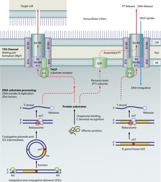
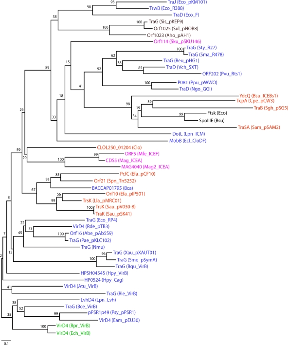
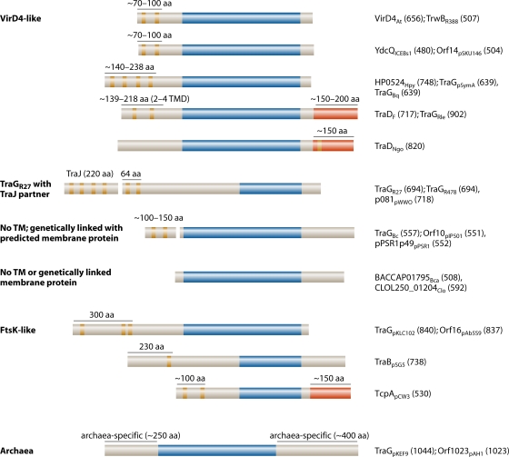
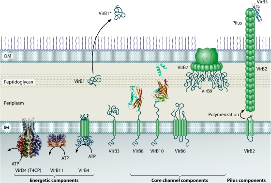
Type IV secretion systems (T4SS) translocate DNA and protein substrates across prokaryotic cell envelopes generally by a mechanism requiring direct contact with a target cell. Three types of T4SS have been described: (i) conjugation systems, operationally defined as machines that translocate DNA substrates intercellularly by a contact-dependent process; (ii) effector translocator systems, functioning to deliver proteins or other macromolecules to eukaryotic target cells; and (iii) DNA release/uptake systems, which translocate DNA to or from the extracellular milieu. Studies of a few paradigmatic systems, notably the conjugation systems of plasmids F, R388, RP4, and pKM101 and the Agrobacterium tumefaciens VirB/VirD4 system, have supplied important insights into the structure, function, and mechanism of action of type IV secretion machines. Information on these systems is updated, with emphasis on recent exciting structural advances. An underappreciated feature of T4SS, most notably of the conjugation subfamily, is that they are widely distributed among many species of gram-negative and -positive bacteria, wall-less bacteria, and the Archaea. Conjugation-mediated lateral gene transfer has shaped the genomes of most if not all prokaryotes over evolutionary time and also contributed in the short term to the dissemination of antibiotic resistance and other virulence traits among medically important pathogens. How have these machines adapted to function across envelopes of distantly related microorganisms? A survey of T4SS functioning in phylogenetically diverse species highlights the biological complexity of these translocation systems and identifies common mechanistic themes as well as novel adaptations for specialized purposes relating to the modulation of the donor-target cell interaction.


Figures

FIG. 1.
FIG. 1.
Mechanisms of T4SS. T4SS mediate the contact-dependent transfer of DNA and protein substrates to target cells, and a subset of systems translocates substrates to or from the extracellular milieu. T4SS are comprised of conjugation, effector translocator, and DNA release/uptake subfamilies. Conjugation systems are comprised of conjugative plasmids and ICEs. For conjugative transfer, DNA substrates are processed by (i) excision from the chromosome by excisionase/integrase enzymes or DDE transposases (for ICEs), (ii) processing of the plasmid or ICE circular transfer intermediate at the origin-of-transfer sequence (oriT) by the Dtr factors (the Dtr-oriT complex is termed a relaxosome), (iii) recruitment of the relaxase-T-strand intermediate to the T4CP, and (iv) translocation through the T4SS channel. Alternatively, protein substrates are maintained in a translocation-competent form and delivered to the T4CP or another receptor or translocation system, e.g., GSP, through the binding of secretion chaperones or other adaptors or spatial-positioning factors. In gram-negative bacteria, T4SS can mediate contact-dependent (left route) or -independent (right route) substrate transfer. The substrate transfer pathway (dashed red lines) through the channel is not clear at this time. DNA uptake by the Helicobacter pylori ComB system (blue line) occurs independently of a T4CP; DNA release by the Neisseria gonorrhoeae GGI-encoded system occurs through a conjugation-like mechanism requiring Dtr factors, a T4CP, and a T4SS channel. In the figure, A. tumefaciens VirD4 is representative of the T4CPs, and the VirB subunits are representative of Mpf channel components; other gram-negative bacterial T4SS are composed of a variable number of VirB homologs. OM, outer membrane; IM, inner membrane; Peri, periplasm.
FIG. 2.
FIG. 2.
Phylogenetic tree of T4CP family members. Sequence alignment and phylogeny estimation were performed using the program MAFFT, version 6.0 (available at http://align.bmr.kyushu-u.ac.jp/mafft/software). The sequence alignment was performed by using the E-INS-i method and default parameters of the program. The tree was constructed using the neighbor-joining method and the following parameters: all ungapped sites from the alignment, JTT amino acid substitution model, and ignore heterogeneity among sites. Bootstrap values for 500 replicates are indicated. The different groups of bacteria are indicated by the following color scheme: blue for gram-negative bacteria, green for gram-negative obligate intracellular pathogens, orange for gram-positive bacteria, pink for cell wall-less bacteria, dark red for Archaea, and black for the related DNA translocases FtsK and SpoIIIE. T4CP designations include the following protein names followed by the species name and plasmid, ICE, or T4SS in parentheses, according to the GenBank database. Accession numbers of T4CPs are AAB58711.1 for Eco_pKM101_TraJ, CAA44852.1 for Eco_R388_TrwB, BAA97972 for Eco_F_TraD, YP_138373.1 for Sis_pKEF9_TraG, CAA09120.1 for Sul_pNOB8_Orf1025, ACI15704.1 for Aho_pAH1_Orf1023, AAS59568.1 for Sku_pSKU146_Orf14, NP_058332.1 for Sty_R27_TraG, NP_941281.1 for Sma_R478_TraG, NP_943001.1 for Reu_pHG1_TraG, AAL59680.1 for Vch_SXT_TraD, NP_640162.1 for Pvu_Rts1_ORF202, NP_542873.1 for Ppu_pWWO_p081, AAW83057.1 for Ngo_GGI_TraD, CAB12293.1 for Bsu_ICEBs1_YdcQ, ABF47325.1 for Cpe_pCW3_TcpA, CAA56759.1 for Sgh_pSG5_TraB, CAA90178.1 for Eco_FtsK, NP_389562.1 for Bsu_SpoIIIE, CAA06449.1 for Sam_pSAM2_TraSA, O54524 for Lpn_ICM_DotL, CAB62409.1 for Ecl_CloDF_MobB, ZP_02074434.1 for Clo_CLOL250_01204, AAN85238.1 for Mfe_ICEF_Orf5, CAJ32610.1 for Mag_ICEA_CDS5, CAL59102.1 for Mag2_ICEA_MAG4040, YP_195789.1 for Efa_pCF10_PcfC, AAG38037.1 for Spn_Tn5252_Orf21, ZP_02036195.1 for Bca_BACCAP01795, CAD44390.1 for Efa_pIP501_Orf10, NP_047302.1 for Lla_pMRC01_TrsK, YP_001653098.1 for Sau_pV030-8_TrsK, NP_863634.1 for Sau_pSK41_TraK, CAA38334.1 for Eco_RP4_TraG, YP_771875.1 for Rde_pTB3_VirD4, YP_001220615.1 for Abe_pAb5S9_Orf16, AAP22624.1 for Pae_pKLC102_TraG, YP_413489.1 for Nmu_TraG, YP_001409435.1 for Xau_pXAUT01_TraG, NP_435748.1 for Sme_pSymA_TraG, YP_032630.1 for Bqu_VirB_TraG, YP_001910374.1 for Hpy_VirB_HPSH04545, NP_207320.1 for Hpy_Cag_HP0524, AAF77174.1 for Atu_VirB_VirD4, ZP_02859218.1 for Rle_VirB_TraG, CAB60062.1 for Lpn_Lvh_LvhD4, ABF79722.1 for Bce_VirB_TraG, NP_940734.1 for Psy_pPSR1_pPSR1p49, NP_943287.1 for Eam_pEU30_VirD4, H71684 for Rpr_VirB_VirD4, and Q8RPL9 for Ech_VirB_VirD4. Species name abbreviations are as follows: Eco, E. coli; Sis, Sulfolobus islandicus; Sul, Sulfolobus sp. strain NOB8H2; Aho, “Acidianus hospitalis”; Sku, S. kunkelii; Sty, S. enterica serovar Typhi; Sma, Serratia marcescens; Reu, Ralstonia eutropha; Vch, V. cholerae; Pvu, Proteus vulgaris; Ppu, Pseudomonas putida; Ngo, N. gonorrhoeae; Bsu, B. subtilis; Cpe, C. perfringens; Sgh, Streptomyces ghanaensis; Sam, Streptomyces ambofaciens; Lpn, L. pneumophila; Eclo, Enterobacter cloacae; Clo, Clostridium sp. strain L2-50; Mfe, M. fermentans; Mag, M. agalactiae strain 5632; Mag2, M. agalactiae PG2; Efa, E. faecalis; Spn, S. pneumoniae; Bca, B. capillosus; Lla, L. lactis; Sau, S. aureus; Rde, Roseobacter denitrificans; Abe, Aeromonas bestiarum; Pae, Pseudomonas aeruginosa; Nmu, Nitrosospira multiformis; Xau, Xanthobacter autotrophicus; Sme, Sinorhizobium meliloti; Bqu, Bartonella quintana; Hpy, H. pylori; Atu, A. tumefaciens; Rle, Rhizobium leguminosarum; Bce, B. cenocepacia; Psy, P. syringae; Eam, Erwinia amylovora; Rpr, R. prowazekii; Ech, E. chaffeensis.
FIG. 3.
FIG. 3.
Distinct molecular architectures of T4CP family members. T4CPs possess recognizable P-loop NTP binding domains but display considerable variation in NTDs and CTDs. The different types of T4CPs are listed at the left, with representatives of each type at the right. T4CPs are designated with the protein name or accession number, with a subscript identifying the species or plasmid origin, followed by the length in amino acid (aa) residues in parentheses. The domains are indicated with the following color scheme: tan, predicted TM helices; dark blue, P-loop domains; red, C-terminal extensions that are ancestrally unrelated to C termini of other T4CPs. Protein sequence analyses were performed by using the programs TMHMM (TM helix prediction), BLASTP (local alignment), and the NCBI conserved-domain search tool. Protein sequences were obtained from the GenBank database (NCBI), and accession numbers are presented in the legend of Fig. 2. TMD, TM domain.
FIG. 4.
FIG. 4.
Localization of the A. tumefaciens VirB/VirD4 subunits. The coupling protein VirD4 and the Mpf components (VirB1 to VirB11) are represented according to their proposed functions: energetic, channel, or pilus components. IM, inner membrane; OM, outer membrane. VirB1 is processed to form VirB1*, which is exported across the outer membrane. VirB2 undergoes a novel head-to-tail cyclization reaction and polymerizes to form the T pilus. A VirB7 lipoprotein-VirB9-VirB10 complex forms a multimeric channel across the outer membrane. Crystal structures are shown for homologs of VirD4 (TrwBR388 soluble domain with an N-terminal TM domain modeled from electron microscopy images) (adapted from reference by permission of Macmillan Publishers Ltd., copyright 2001), VirB11 (HP0525Hp) (adapted from reference with permission of Elsevier), VirB5 (TraCpKM101) (adapted from reference with permission of the publisher; copyright 2005 National Academy of Sciences, U.S.A.), VirB8 (soluble domain of VirB8Bs), and VirB10 (soluble domain of ComB10Hp) (VirB8 and VirB10 adapted from reference with permission of the publisher. Copyright 2003 National Academy of Sciences, U.S.A.). VirB7, VirB9, and VirB10 assemble to form a transenvelope “core” complex (Fig. 5).
FIG. 5.
FIG. 5.
Substrate translocation pathway and architecture of a T4SS core complex. The substrate translocation pathway depicted at the left (red arrow) was developed for the A. tumefaciens VirB/VirD4 system on the basis of DNA-channel subunit contacts identified by formaldehyde cross-linking (49). The DNA substrate cross-links with the VirD4 T4CP, VirB11 ATPase, polytopic VirB6, bitopic VirB8, VirB2 pilin, and outer membrane (OM) VirB9. These subunits are postulated to comprise the secretion channel, whereas other components, including VirB3, VirB4, and VirB10, promote channel assembly as protein scaffolds or through ATP-mediated conformational changes. A CryoEM structure of a core complex composed of pKM101 VirB7-like TraN, VirB9-like TraO, and VirB10-like TraF is presented at right (102). Fourteen copies of each subunit assemble to form a large double-walled chamber that presumptively spans the entire gram-negative cell envelope. Additional subunits listed at the entrance are postulated to position within the core chamber. (The cryo-electron microscopy structure is reprinted from reference with permission from AAAS.)
FIG. 6.
FIG. 6.
T4SS genes in the genomes of gram-negative obligate intracellular pathogens are organized in distinct clusters. (A) Organizations of T4SS genes in some completed genomes of intracellular pathogens; noncontiguous gene clusters are separated by double cross-hatched lines (106, 136, 199). All genomes examined possess multiple copies of virB6-like genes. (B) Anaplasmataceae genomes also possess several copies of virB2-like genes often next to virB4-like genes. GenBank accession numbers are CP000235 for A. phagocytophilum, CP000236 for E. chaffeensis, CP000237 for N. sennetsu, and AE017196 for Wolbachia pipientis ωMel. Orthologous and paralogous genes are shown in the same color for all the genomes; gene sizes are not depicted to scale.
FIG. 7.
FIG. 7.
Predicted subunits of five representative T4SS functioning in gram-positive bacteria. Dispositions of the T4SS subunits at the cell envelope are depicted schematically; topology predictions were derived from the TMHMM algorithm and have not been experimentally confirmed. ORFs 9 and 10 from pIP501 might comprise a novel two-partner T4CP, and the VirB4-like proteins might form a complex with an adjacent gene product, as indicated with double arrows and “?.” The VirB6-like subunits are subgrouped as VirB6 (PrgH and Orf12) or extended VirB6 (YddG, TcpH, and Orf15) (see the text for details). Proteins with topologies and/or domains indicative of a role as a T4SS component are depicted, with the exception of TcpIpCW3, a possible membrane-bound metal hydrolase important for pCW3 transfer. Proteins of similar sizes and predicted membrane topologies (TMHMM algorithm) are listed with the same color, and proteins exhibiting sequence relatedness in local alignments using the BLASTP or Psi-BLAST algorithm are listed with the same color and an asterisk. A minus sign indicates that no corresponding subunit is produced by the transfer system. CW, cell wall; CM, cytoplasmic membrane. GenBank accession numbers are NC_006827 for E. faecalis pCF10, L39769.1 and AJ505823.1 for S. agalactiae pIP501, AB001488.1 for B. subtilis ICEBs1, NC_010937.1 for C. perfringens pCW3, and NC_006372.1 for E. faecalis Tn916.
FIG. 8.
FIG. 8.
Surface proteins shown or predicted to be associated with T4SS. (Left) Proteins are grouped according to their sequence relatedness to A. tumefaciens VirB subunits. (Center) Schematics of novel features and domains. SP, signal peptide; lipo box, lipoprotein motif; c-coil, coiled coil; anchor, cell wall anchor motif; aa, amino acids. Gene (arrows, top) and protein (rectangles) sizes are not to scale. (Right) Description of surface variation and possible function. Studies describing demonstrated or postulated functions in the environmental response and/or adaptation are cited. PMN, polymorphonuclear leukocytes; LTA, lipoteichoic acid.

References

    1. Abajy, M. Y., J. Kopec, K. Schiwon, M. Burzynski, M. Doring, C. Bohn, and E. Grohmann. 2007. A type IV-secretion-like system is required for conjugative DNA transport of broad-host-range plasmid pIP501 in gram-positive bacteria. J. Bacteriol. 189:2487-2496. - PMC - PubMed
    1. Abdallah, A. M., N. C. G. van Pittius, P. A. Champion, J. Cox, J. Luirink, C. M. Vandenbroucke-Grauls, B. J. Appelmelk, and W. Bitter. 2007. Type VII secretion—mycobacteria show the way. Nat. Rev. Microbiol. 5:883-891. - PubMed
    1. Al-Khodor, S., C. T. Price, F. Habyarimana, A. Kalia, and Y. Abu Kwaik. 2008. A Dot/Icm-translocated ankyrin protein of Legionella pneumophila is required for intracellular proliferation within human macrophages and protozoa. Mol. Microbiol. 70:908-923. - PMC - PubMed
    1. Aly, K. A., and C. Baron. 2007. The VirB5 protein localizes to the T-pilus tips in Agrobacterium tumefaciens. Microbiology 153:3766-3775. - PubMed
    1. Amor, J. C., J. Swails, X. Zhu, C. R. Roy, H. Nagai, A. Ingmundson, X. Cheng, and R. A. Kahn. 2005. The structure of RalF, an ADP-ribosylation factor guanine nucleotide exchange factor from Legionella pneumophila, reveals the presence of a cap over the active site. J. Biol. Chem. 280:1392-1400. - PubMed

Publication types

LinkOut - more resources