. 2010 Feb 5;285(6):4110-4121.
doi: 10.1074/jbc.M109.073676. Epub 2009 Nov 29.

Dnmt3 and G9a cooperate for tissue-specific development in zebrafish

Affiliations

Dnmt3 and G9a cooperate for tissue-specific development in zebrafish

Kunal Rai et al. J Biol Chem. .

Abstract

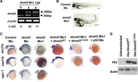
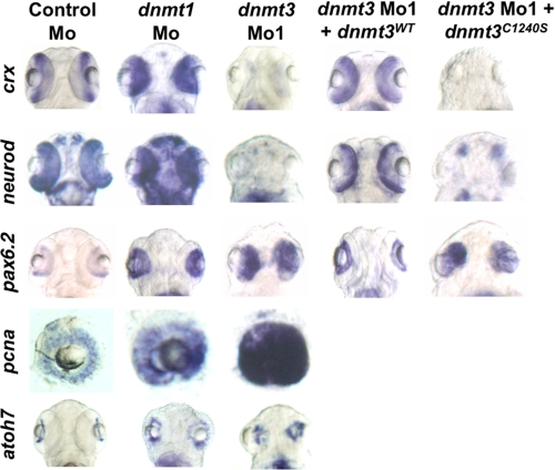
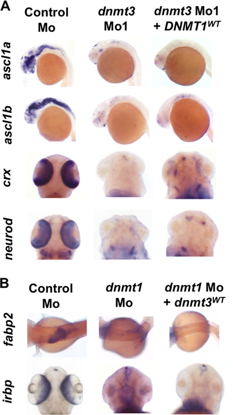
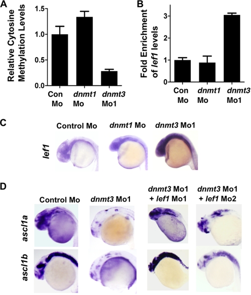
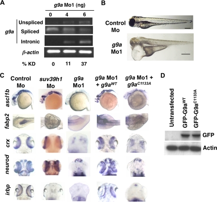
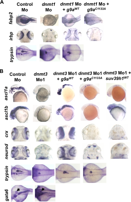
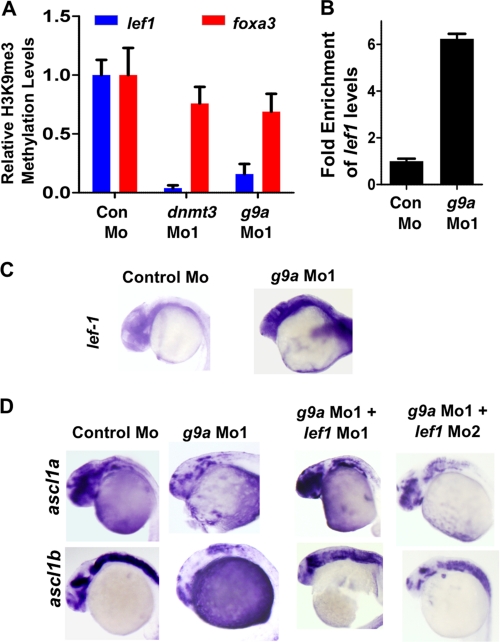
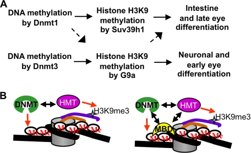
Although DNA methylation is critical for proper embryonic and tissue-specific development, how different DNA methyltransferases affect tissue-specific development and their targets remains unknown. We address this issue in zebrafish through antisense-based morpholino knockdown of Dnmt3 and Dnmt1. Our data reveal that Dnmt3 is required for proper neurogenesis, and its absence results in profound defects in brain and retina. Interestingly, other organs such as intestine remain unaffected suggesting tissue-specific requirements of Dnmt3. Further, comparison of Dnmt1 knockdown phenotypes with those of Dnmt3 suggested that these two families have distinct functions. Consistent with this idea, Dnmt1 failed to complement Dnmt3 deficiency, and Dnmt3 failed to complement Dnmt1 deficiency. Downstream of Dnmt3 we identify a neurogenesis regulator, lef1, as a Dnmt3-specific target gene that is demethylated and up-regulated in dnmt3 morphants. Knockdown of lef1 rescued neurogenesis defects resulting from Dnmt3 absence. Mechanistically, we show cooperation between Dnmt3 and an H3K9 methyltransferase G9a in regulating lef1. Further, like Dnmt1-Suv39h1 cooperativity, Dnmt3 and G9a seemed to function together for tissue-specific development. G9a knockdown, but not Suv39h1 loss, phenocopied dnmt3 morphants and G9a overexpression provided a striking rescue of dnmt3 morphant phenotypes, whereas Suv39h1 overexpression failed, supporting the notion of specific DNMT-histone methyltransferase networks. Consistent with this model, H3K9me3 levels on the lef1 promoter were reduced in both dnmt3 and g9a morphants, and its knockdown rescued neurogenesis defects in g9a morphants. We propose a model wherein specific DNMT-histone methyltransferase networks are utilized to silence critical regulators of cell fate in a tissue-specific manner.


Figures

FIGURE 1.
FIGURE 1.
Dnmt3 knockdown in zebrafish embryos results in neurogenesis defects. A, splice blocking by dnmt3 splice-blocking morpholino (dnmt3 Mo1) was monitored by RT-PCR in mRNAs from control and dnmt3 morphants at 80 hpf. PCR was performed using a forward primer in exon 6 and a reverse primer in exon 7. Note that the dnmt3 Mo1 stabilizes the unspliced transcript containing introns 6 and 7 (400 bp), whereas the spliced version (300 bp) is detected in control injected embryos. Percent knockdown (% KD) shown is the ratio of intensity of unspliced product to combined intensity of unspliced and spliced products normalized over β-actin intensity. B, morphology defects in dnmt3 morphants at 80 hpf. Note the smaller head, a pericardial edema, and a curled tail in dnmt3 morphants compared with control morphants. Bar equals 0.5 mm. C, whole mount in situ analysis of ngn-1, ascl1a, and ascl1b expression in dnmt3 morpholino alone or with p53 morpholino-, dnmt1 morpholino-, or control morpholino-injected embryos at 30 hpf. Whereas ngn-1 expression was normal in dnmt3 and dnmt1 morphants, ascl1a and ascl1b were selectively is absent. This defect can be complemented by co-injection of the wild-type (Dnmt3WT) but not by a catalytically inactive derivative (Dnmt3C1240S). D, protein expression levels of Dnmt3 wild-type (Dnmt3WT) and catalytically inactive (Dnmt3C1240S) derivatives. HEK293 cells were transfected with Dnmt3WT and Dnmt3C1240S, and Western blots were performed using antibodies against His tag. Both of these show equal expression of these derivatives. β-Actin was used as a loading control.
FIGURE 2.
FIGURE 2.
Distinct tissue-specific developmental defects in dnmt1 and dnmt3 morphants. A, C, and E, whole mount in situ analysis of dhand (at 48 hpf) (A), fabp2, trypsin, fabp10, insulin, and trypsin (C), and irbp (E) (all at 96 hpf) in dnmt1, dnmt3, and control morphants. In panel A, arrows show loss of last two pharyngeal pouches in dnmt3 morphants. In panel C, arrows point to expression of gata6 in the pancreas. B, Alcian Blue staining of Control, dnmt1, or dnmt3 morphants at 96 hpf shows loss of jaw structure in dnmt1 morphants but not in dnmt3 morphants. D, toluidine blue staining of histological cross-sections within the dnmt3 morphant retinas at 80 hpf. Note the loss of organization of different retinal layers in dnmt3 morphants. F, quantitative RT-PCR for Opsin Shortwave 1 and Opsin Shortwave 2 in control, dnmt1, or dnmt3 morpholino-injected embryos at 96 hpf.
FIGURE 3.
FIGURE 3.
Dnmt3 morphants are defective in retinal differentiation. Whole mount in situ for pax6.2, crx, and neurod in dnmt3 and control morphants at 80 hpf. Whereas all the retinal cells expressed pax6.2, a marker of retinal progenitors, markers of specific progenitors (crx and neurod) were absent in dnmt3 morphants. These defects can be complemented by co-injection of the wild-type Dnmt3 (Dnmt3WT) but not with the catalytically inactive Dnmt3 derivative (Dnmt3C1240S).
FIGURE 4.
FIGURE 4.
Dnmt3 overexpression in dnmt1 morphants and Dnmt1 overexpression in dnmt3 morphants does not rescue developmental defects. A and B, whole mount in situ expression for ascl1a, ascl1b, crx, and neurod (A) and fabp2 and irbp (B) in embryos injected with control morpholino, dnmt3 morpholino alone, or with DNMT1 mRNA (A) and dnmt1 morpholino alone or with dnmt3 mRNA (B).
FIGURE 5.
FIGURE 5.
Lef1 repression by Dnmt3 is critical for proper neurogenesis. A, MeDIP-PCR based quantification of methylation status of lef1 promoter in control, dnmt1, and dnmt3 morphant embryos at 24 hpf. The y-axis represents values at lef1 promoter first normalized to a negative region (with an insignificant number of CpGs in 1000 bp vicinity) and then normalized to control Mo values as 1. B, graph showing quantitative RT-PCR results for zebrafish lef1 normalized to 28 S values in control morpholino-, dnmt1 morpholino-, or dnmt3 morpholino-injected embryos at 24 hpf. Results are represented in a -fold change format where the lef1/28S ratio from control morphants was normalized to 1. C, expression of lef1 in control, dnmt1, and dnmt3 morphant embryos at 24 hpf as detected by whole mount in situ hybridization. D, whole mount in situ analysis of ascl1a and ascl1b expression in embryos (30 hpf) injected with control morpholino and dnmt3 morpholino co-injected with either lef1 Mo1 or lef1 Mo2.
FIGURE 6.
FIGURE 6.
G9a morphants largely phenocopy dnmt3 morphants. A, splice blocking by g9a splice-blocking morpholino (g9a Mo1) was monitored by RT-PCR in mRNAs made from control and g9a morphants at 80 hpf using a forward primer in the exon and reverse primer in the next exon. Note that the g9a morpholino stabilizes the unspliced transcript containing the intermediate intron, whereas the spliced version is detected in control-injected embryos. The percent knockdown shown is the ratio of intensity of unspliced product to combined intensity of unspliced and spliced products normalized to intensity of the β-actin band. Note that a product can be amplified with an intronic primer in the g9a morphants; however, this was not taken into account while calculating percent knockdown. B, morphology defects in g9a morphants at 80 hpf. Note the smaller head in g9a morphants, as with dnmt3 morphants. Bar equals 0.5 mm. C, whole mount in situ analysis of ascl1b, crx, neurod, fabp2, and irbp expression in g9a, suv39h1, and control morphants. ascl1b was analyzed at 30 hpf, whereas all others were analyzed at 80 hpf. The defects present in g9a morphants were complemented by co-injection of the wild-type G9a (G9aWT), but not by the catalytically inactive derivative G9aC1133A. D, expression of exogenous G9a constructs. HEK293 cells were transfected with the wild-type or catalytically dead derivatives of GFP-tagged zebrafish G9a (G9aWT and G9aC1133A). Westerns using antibodies against the GFP tag show equal expression of these derivatives. β-Actin was used a loading control.
FIGURE 7.
FIGURE 7.
G9a overexpression rescues both dnmt3 morphants and dnmt1 morphants. Whole mount in situ analysis of ascl1a, ascl1b, crx, and neurod (A) and fabp2, irbp, and trypsin (B) expression in embryos injected with control morpholino or dnmt3 or dnmt1 morpholino co-injected with wild-type or catalytically null G9a or Suv39h1. ascl1a and ascl1b were analyzed at 30 hpf, whereas all others were analyzed at 80 hpf. Note that co-injection of wild-type G9a (G9aWT) but not of either catalytically inactive G9a (G9aC1133S) or wild-type Suv39h1 rescues the ascl1a and ascl1b expression in dnmt3 morphants. Overexpression of wild-type G9a, but not catalytically inactive G9a, can also rescue fabp2 and irbp expression in dnmt1 morphants. Of note, wild-type G9a could not rescue trypsin expression in dnmt1 morphants. The panel showing Suv39h1 rescue of dnmt1 morphants has been reported previously (7) and is shown for comparative purposes.
FIGURE 8.
FIGURE 8.
Lef1 repression by G9a is critical for proper neurogenesis. A, quantitative PCR for chromatin immunoprecipitation for H3K9me3 marks on lef1 promoter (Blue) or on foxa3 promoter (Red) in control morphant, dnmt3 morphant, and g9a morphant embryos. Values shown represent enrichment on the experimental region normalized to values on control region. B, graph showing quantitative RT-PCR results for zebrafish lef1 normalized to 28S values in control morpholino or g9a morpholino-injected embryos at 24 hpf. Results are represented in a -fold change format where the lef1/28S ratio from control morphants was normalized to 1. C, expression of lef1 in control and g9a morphant embryos at 24 hpf as detected by whole mount in situ hybridization. D, whole mount in situ analysis of ascl1a and ascl1b expression in embryos (30 hpf) injected with control morpholino and g9a morpholino co-injected with either lef1 Mo1 or lef1 Mo2.
FIGURE 9.
FIGURE 9.
Models for DNMT-HMT suppression relationships and cooperativity. A, DNMTs methylate the cytosines in the CpG island present in the promoter of the critical regulator of differentiation (for example, Lef1), which is required to be silenced to promote differentiation. DNA methylation marks lead to recruitment of histone H3K9 methyltransferases (HMTs), which then mark the histone tails to help establish a repressed state. Different DNMTs recruit different HMTs; Dnmt3 depends specifically on G9a, whereas Dnmt1 can recruit both Suv39h1 and G9a (but relies more on Suv39h1). This specific DNMT-HMT network is responsible for promoting specific differentiation stages in different organs. B, two physical models can be proposed for the dependence of a DNMT on an HMT. First (left panel), a DNA methyltransferase could directly recruit specific HMT on the promoter of a given gene, and this specific interaction between DNMT and HMT is sufficient to generate specificity. Second (right panel), there exists a ternary complex composed of a DNMT, a methyl binding domain-containing protein (MBD), and an HMT. Specific interactions between these three proteins govern specificity toward their gene targets. Initial recruitment may rely on gene-specific DNA-binding proteins (not shown).

Similar articles

Cited by

References

    1. Razin A., Cedar H. (1991) Microbiol. Rev. 55, 451–458 - PMC - PubMed
    1. Bird A. P. (1986) Nature 321, 209–213 - PubMed
    1. Rai K., Nadauld L. D., Chidester S., Manos E. J., James S. R., Karpf A. R., Cairns B. R., Jones D. A. (2006) Mol. Cell. Biol. 26, 7077–7085 - PMC - PubMed
    1. Anderson R. M., Bosch J. A., Goll M. G., Hesselson D., Dong P. D., Shin D., Chi N. C., Shin C. H., Schlegel A., Halpern M., Stainier D. Y. (2009) Dev. Biol. 334, 213–223 - PMC - PubMed
    1. Goll M. G., Bestor T. H. (2005) Annu. Rev. Biochem. 74, 481–514 - PubMed

Publication types

MeSH terms

Substances