

A long DNA segment in a linear nanoscale Paul trap
- PMID: 19946172
- PMCID: PMC3269948
- DOI: 10.1088/0957-4484/21/1/015103
A long DNA segment in a linear nanoscale Paul trap
Abstract
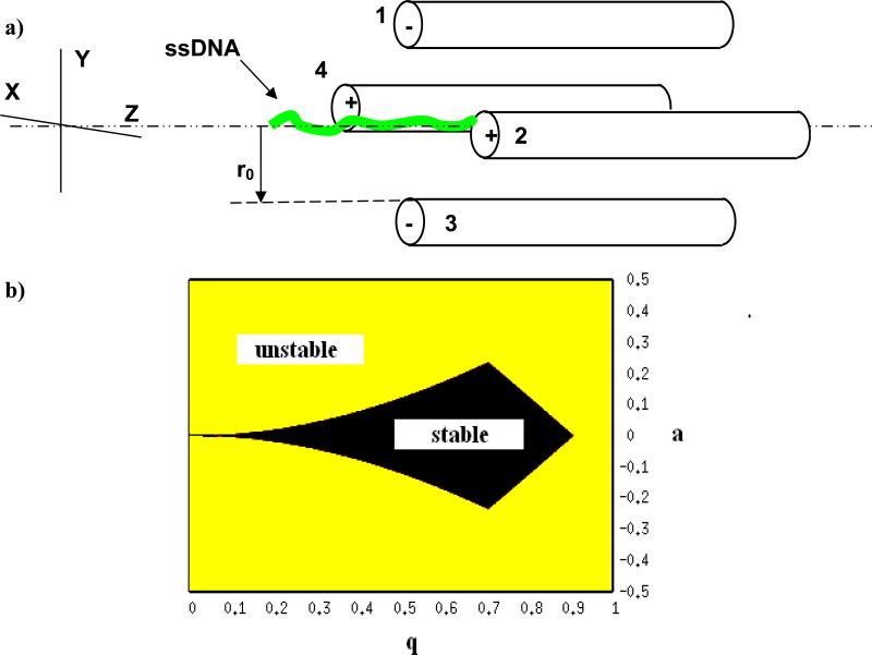
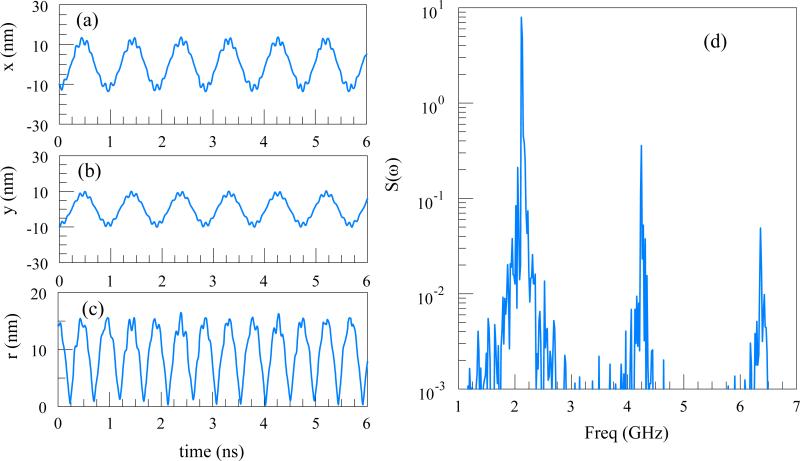
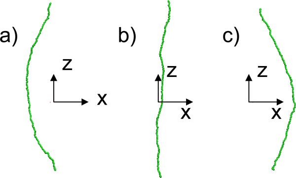
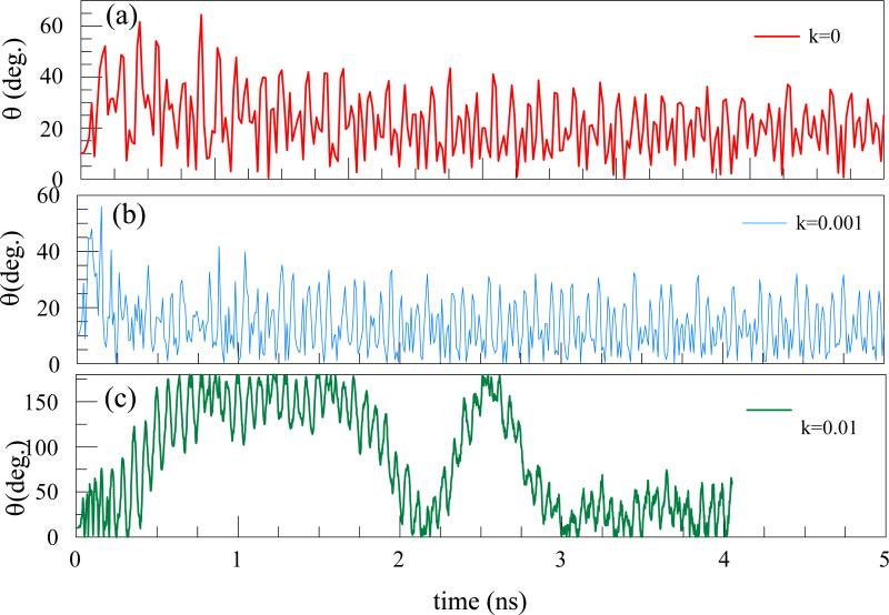
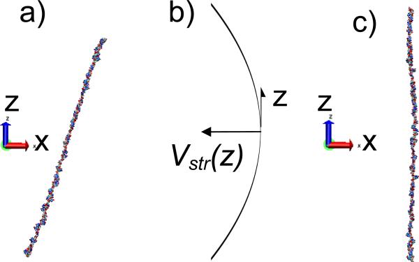
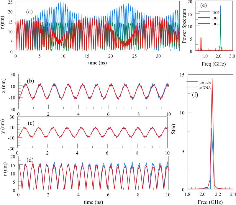
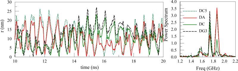
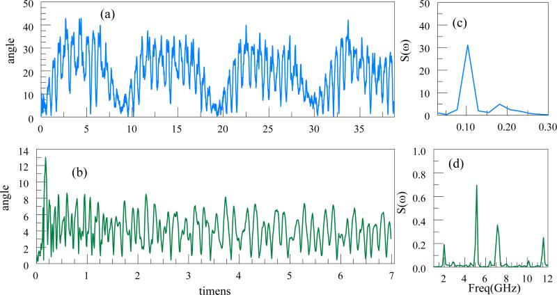
We study the dynamics of a linearly distributed line charge such as single stranded DNA (ssDNA) in a nanoscale, linear 2D Paul trap in vacuum. Using molecular dynamics simulations we show that a line charge can be trapped effectively in the trap for a well defined range of stability parameters. We investigated (i) a flexible bonded string of charged beads and (ii) a ssDNA polymer of variable length, for various trap parameters. A line charge undergoes oscillations or rotations as it moves, depending on its initial angle, the position of the center of mass and the velocity. The stability region for a strongly bonded line of charged beads is similar to that of a single ion with the same charge to mass ratio. Single stranded DNA as long as 40 nm does not fold or curl in the Paul trap, but could undergo rotations about the center of mass. However, we show that a stretching field in the axial direction can effectively prevent the rotations and increase the confinement stability.
Figures


Similar articles
-
Control Of Screening Of A Charged Particle In Electrolytic Aqueous Paul Trap.AIP Conf Proc. 2011;1336:150-153. doi: 10.1063/1.3586077. AIP Conf Proc. 2011. PMID: 24839332 Free PMC article.
-
A molecular dynamics simulation study on trapping ions in a nanoscale Paul trap.Nanotechnology. 2008 May 14;19(19):195702. doi: 10.1088/0957-4484/19/19/195702. Epub 2008 Apr 8. Nanotechnology. 2008. PMID: 21825720 Free PMC article.
-
Effects of Polymer Length and Salt Concentration on the Transport of ssDNA in Nanofluidic Channels.Biophys J. 2017 Mar 14;112(5):838-849. doi: 10.1016/j.bpj.2017.01.027. Biophys J. 2017. PMID: 28297643 Free PMC article.
-
Molecular dynamics simulation of ss-DNA translocation between copper nanoelectrodes incorporating electrode charge dynamics.J Phys Chem B. 2008 Feb 14;112(6):1712-7. doi: 10.1021/jp077483e. Epub 2008 Jan 23. J Phys Chem B. 2008. PMID: 18211061
-
Nanoscale mechanical and dynamical properties of DNA single molecules.Biophys Chem. 2005 Mar 1;113(3):209-21. doi: 10.1016/j.bpc.2004.09.007. Biophys Chem. 2005. PMID: 15620506 Review.
Cited by
-
Paul trapping of charged particles in aqueous solution.Proc Natl Acad Sci U S A. 2011 Jun 7;108(23):9326-30. doi: 10.1073/pnas.1100977108. Epub 2011 May 23. Proc Natl Acad Sci U S A. 2011. PMID: 21606331 Free PMC article.
-
Control and reversal of the electrophoretic force on DNA in a charged nanopore.J Phys Condens Matter. 2010 Nov 17;22(45):454123. doi: 10.1088/0953-8984/22/45/454123. Epub 2010 Oct 29. J Phys Condens Matter. 2010. PMID: 21339610 Free PMC article.
-
Control Of Screening Of A Charged Particle In Electrolytic Aqueous Paul Trap.AIP Conf Proc. 2011;1336:150-153. doi: 10.1063/1.3586077. AIP Conf Proc. 2011. PMID: 24839332 Free PMC article.
-
Thermal noise in aqueous quadrupole micro- and nano-traps.Nanoscale Res Lett. 2012 Feb 27;7(1):156. doi: 10.1186/1556-276X-7-156. Nanoscale Res Lett. 2012. PMID: 22369362 Free PMC article.
-
Stability of an aqueous quadrupole micro-trap.J Phys Condens Matter. 2012 Apr 25;24(16):164208. doi: 10.1088/0953-8984/24/16/164208. Epub 2012 Mar 30. J Phys Condens Matter. 2012. PMID: 22466254 Free PMC article.
References
-
- Abich K, Keil A, Reiss D, Wunderlich C, Neuhauser W, Toschek PE. Thermally activated hopping of two ions trapped in a bistable potential well. Journal of Optics B-Quantum and Semiclassical Optics. 2004;6:S18–S23.
-
- Aksimentiev A, Schulten K, Heng J, Ho C, Timp G. Molecular dynamics simulations of a nanopore device for DNA sequencing. Biophysical Journal. 2004b;86:480A–A.
-
- Arnott D, Henzel WJ, Stults JT. Rapid identification of comigrating gel-isolated proteins by ion trap mass spectrometry. Electrophoresis. 1998;19:968–80. - PubMed
Publication types
MeSH terms
Substances
Grants and funding
LinkOut - more resources
Full Text Sources
Other Literature Sources