. 2009 Dec 24;462(7276):1011-5.
doi: 10.1038/nature08588. Epub 2009 Nov 29.

Structure of the outer membrane complex of a type IV secretion system

Affiliations

Structure of the outer membrane complex of a type IV secretion system

Vidya Chandran et al. Nature. .

Abstract

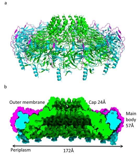
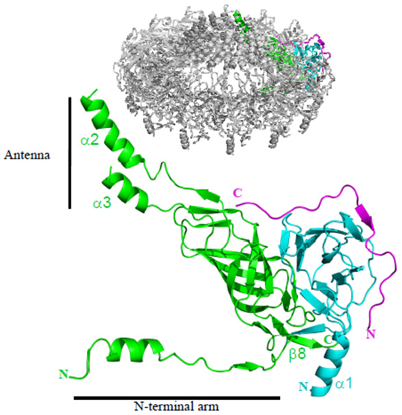
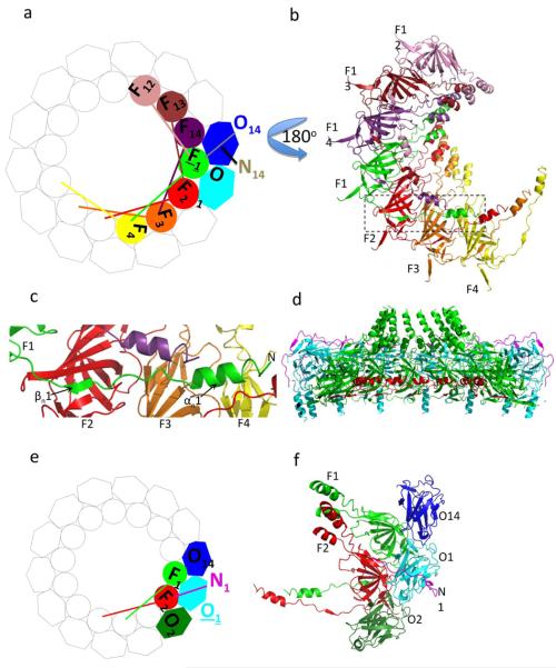
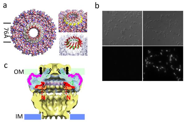
Type IV secretion systems are secretion nanomachines spanning the two membranes of Gram-negative bacteria. Three proteins, VirB7, VirB9 and VirB10, assemble into a 1.05 megadalton (MDa) core spanning the inner and outer membranes. This core consists of 14 copies of each of the proteins and forms two layers, the I and O layers, inserting in the inner and outer membrane, respectively. Here we present the crystal structure of a approximately 0.6 MDa outer-membrane complex containing the entire O layer. This structure is the largest determined for an outer-membrane channel and is unprecedented in being composed of three proteins. Unexpectedly, this structure identifies VirB10 as the outer-membrane channel with a unique hydrophobic double-helical transmembrane region. This structure establishes VirB10 as the only known protein crossing both membranes of Gram-negative bacteria. Comparison of the cryo-electron microscopy (cryo-EM) and crystallographic structures points to conformational changes regulating channel opening and closing.


Figures

Figure 1
Figure 1
The T4S system outer membrane complex. Ribbon diagram (a) and space-filling cut-away (b) of the tetradecameric complex. TraFCT, TraOCT and TraN subunits are colour-coded green, cyan, and magenta, respectively. In b, dimensions and labelling of the various parts of the complex are provided.
Figure 2
Figure 2
Ribbon diagram of the heterotrimer unit. Structures mentioned in the main text are indicated. The insert locates the shown heterotrimer within the tetradecameric structure. A stereo version of this figure with full secondary structure labelling is provided in Supplementary Fig. 2d.
Figure 3
Figure 3
Inter-heterotrimeric interactions. a. Schematic diagram of the tetradecamer with emphasis on interactions with the TraFCT subunit in green (F1). TraFCT and TraOCT subunits are shown in circles and hexagons, respectively. Not shown for clarity: TraN subunits (except N14 that makes interactions with F1) and the lever arms of subunits not interacting with F1. b. Ribbon diagram of the F1-interacting TraFCT subunits (i.e F4, F3, F2, F14, F13, and F12) viewed from the periplasm i.e. turned 180° compared to the view in panel a. The rectangle locates the lever arm of subunit F1. c. Interactions of subunits F2, F3, and F4 with the lever arm of subunit F1. The secondary structures of the lever arm of F1 are labelled. The F2, F3, and F4 subunits are in ribbon representation and labelled accordingly. d. Cut-away side view of the outer membrane complex with the proteins in ribbon diagram representation color-coded as in Fig. 1 but for the N-terminal arms of the TraFCT subunits in red. e. Schematic diagram of the tetradecamer with emphasis on interactions with the TraOCT subunit in cyan (O1). Not shown for clarity: TraN subunits (except N1 that makes interactions with O1) and the lever arms of subunits not interacting with O1. f. Ribbon diagram of the subunits shown in colour at left. This view is from the extracellular milieu.
Figure 4
Figure 4
Trans-membrane region of the T4S system outer membrane complex and proposed mechanism of pore opening/closure. a. Left panel: surface diagram of complex viewed from the extracellular milieu. The two lines define the hydrophobic central region around the pore. Top right panel: same as at left but showing (in yellow) the ring of Trp residues at the base of α2. Bottom right panel: ribbons diagram of the trans-membrane helices with the internal and external ring of helices colour-coded in green and red, respectively. b. Extracellular detection of the FLAG-tag located between helices α2 and α3 of TraF. A FLAG-tag was introduced in the loop between the α2 and α3 helices of TraF as described in methods. Upper and lower left panels: control sample expressing the wild-type T4SS core complex. Upper and lower right panels: cells expressing the FLAG-tagged T4SS core complex. Differential interference contrast images are shown in the upper panels and the corresponding fluorescence images in the lower panels. c. Proposed mechanism for pore opening and closure. The cryo-EM core complex structure (pale yellow) is superimposed on the crystal structure of the outer membrane complex (pale cyan). The N-terminal lever arm shelf is highlighted in orange and red in the EM and crystal structures, respectively. The position of the lipid on TraN/VirB7 is indicated in black dots. TraN/VirB7 is shown in magenta. Proposed conformational changes are illustrated by arrows. The N-terminal sequences linking the lever arms to sequences in the I-layer are shown in dashed lines colour-coded red and orange for the conformations found in the crystal and EM structures, respectively.

Comment in

References

    1. Cascales E, Christie PJ. The versatile bacterial type IV secretion systems. Nat Rev Microbiol. 2003;1:137–149. - PMC - PubMed
    1. Christie PJ, Atmakuri K, Krishnamoorthy V, Jakubowski S, Cascales E. Biogenesis, architecture, and function of bacterial type IV secretion systems. Annu Rev Microbiol. 2005;59:451–485. - PMC - PubMed
    1. Schroder G, Lanka E. The mating pair formation system of conjugative plasmids-A versatile secretion machinery for transfer of proteins and DNA. Plasmid. 2005;54:1–25. - PubMed
    1. Backert S, Selbach M. Role of type IV secretion in Helicobacter pylori pathogenesis. Cell Microbiol. 2008;10:1573–1581. - PubMed
    1. Ninio S, Roy CR. Effector proteins translocated by Legionella pneumophila: strength in numbers. Trends Microbiol. 2007;15:372–380. - PubMed

References for Methods section

    1. Sharff AJ, Koronakis E, Luisi B, Koronakis V. Oxidation of selenomethionine: some MADness in the method! Acta Crystallogr D Biological Crystallography. 2000;56:785–788. - PubMed
    1. Kabsch W. Automatic processing of rotation diffraction data from crystals of initially unknown symmetry and cell constants. Journal of applied crystallography. 1993;26:795–800.
    1. Navaza J. AMoRe: an automated package for molecular replacement. Acta Crystallographica Section A: Foundations of Crystallography. 1994;50:157–163.
    1. Jones TA. A set of averaging programs. In "Molecular Replacement". CCP4 Proceedings. 1992:91–95.
    1. Kjeldgaard M, Jones TA. Halloween … masks and bones. In “From First Map to Final Model”. CCP4 Proceedings. 1994:59–66.

Publication types

MeSH terms

Substances

Associated data