. 2009 Dec 17;462(7275):920-4.
doi: 10.1038/nature08577. Epub 2009 Nov 29.

Stably maintained dendritic spines are associated with lifelong memories

Affiliations

Stably maintained dendritic spines are associated with lifelong memories

Guang Yang et al. Nature. .

Abstract

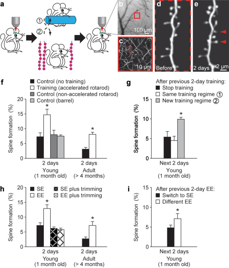
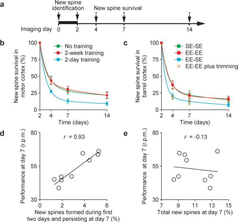
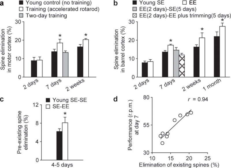
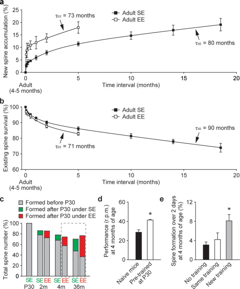
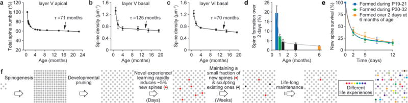
Changes in synaptic connections are considered essential for learning and memory formation. However, it is unknown how neural circuits undergo continuous synaptic changes during learning while maintaining lifelong memories. Here we show, by following postsynaptic dendritic spines over time in the mouse cortex, that learning and novel sensory experience lead to spine formation and elimination by a protracted process. The extent of spine remodelling correlates with behavioural improvement after learning, suggesting a crucial role of synaptic structural plasticity in memory formation. Importantly, a small fraction of new spines induced by novel experience, together with most spines formed early during development and surviving experience-dependent elimination, are preserved and provide a structural basis for memory retention throughout the entire life of an animal. These studies indicate that learning and daily sensory experience leave minute but permanent marks on cortical connections and suggest that lifelong memories are stored in largely stably connected synaptic networks.


Figures

Figure 1
Figure 1. Motor learning and novel sensory experience promote rapid dendritic spine formation
a, Transcranial two-photon imaging of spines before and after rotarod training or sensory enrichment. b, CCD camera view of the vasculature of the motor cortex. c, Two-photon image of apical dendrites from the boxed region in b. A higher-magnification view of a dendritic segment in c is shown in d. d–e, Repeated imaging of a dendritic branch before (d) and after rotarod training (e). Arrowheads indicate new spines formed over 2 days. f, Percentage of new spines formed within 2 days in the motor cortex was significantly higher in young or adult mice after training as compared with controls with no training or running on a non-accelerated rotarod. No increase in spine formation was found in the barrel cortex after training. g, After previous 2-day training, only a new training regime (reverse running) caused a significant increase in spine formation. h, EE increased spine formation over 2 days in the barrel cortex in both young and adult animals. No significant increase in spine formation was found under EE when the whiskers were trimmed. i. After previous 2-day EE, animals switched to a different EE showed a higher rate of spine formation than those returned to SE. Data are presented as mean ± s.d. *P < 0.005. See Supplementary Table for the number of animals in each group.
Figure 2
Figure 2. A fraction of newly formed spines persists over weeks and correlates with performance after learning
a, New spines induced by novel experience were identified in the first 2 days and followed over time. b, c, The survival of new spines (mean ± s.d.) over time under various conditions. A significantly larger fraction of new spines remained in mice trained repeatedly or housed under EE continuously. The lines represent two exponential fittings (r2 = 1). d, e, An animal’s performance at day 7 strongly correlated with new spines formed during the first 2-day training and persisting at day 7 (d), but did not correlate with the total new spines accumulated from day 0 to 7 (e). Each circle represents an individual animal. The linear regression lines and correlation coefficients (r) are shown.
Figure 3
Figure 3. Novel experience promotes spine elimination
a, b, Percentage of spines eliminated (mean ± s.d.) in young animals under various conditions. Rotarod training (a) or EE (b) for at least 7 days increased the elimination of existing spines (P < 0.05). c, EE increased the elimination of spines that existed for more than 2 days before EE exposure (P < 0.05). d, The elimination of existing spines over 7 days strongly correlated with an animal’s performance on day 7 (r = 0.94). Each circle represents an individual animal.
Figure 4
Figure 4. Maintenance of daily formed new spines and spines formed during early development throughout life
a, New spine accumulation over time under SE and EE. Three exponential fits show that ~0.8% of daily formed new spines decay with a time constant of 80 months under SE and 73 months under EE. b, The percentage of adult spines remaining over time under SE and EE. Three exponential fits show that ~90% of adult spines have an average lifetime of 90 months under SE and 71 months under EE. c, A large fraction of spines formed before P30 persisted throughout life under SE or EE. The projections based on a and b are shown in the dashed frame. d, Mice previously trained at P30 for 7 days showed better performance (mean ± s.e.m.) when assessed at 4 months of age than naive mice (P < 0.01). e, Only a new training regime (reverse running) caused an increase in spine formation in previously trained animals. Spine data are presented as mean ± s.d.
Figure 5
Figure 5. Spine maintenance in different cell types and cortical layers
ac, Age-dependent change in spine number is remarkably similar across different cell types/cortical layers in barrel cortex and contains information on spine dynamics. Total spine number (percentage of P19) of layer V pyramidal cell apical dendrites (a) was measured through in vivo imaging. Spine densities (mean ± s.e.m.) of layer V and layer VI pyramidal cell basal dendrites (b, c) were measured on dendritic segments located 50–100 μm from the soma in fixed brain slices. d, Spine formation rate declined rapidly from P19 to P30 and remained low thereafter. e, Regardless of animals’ ages (P19, P30, 6 months), a fraction of new spines formed over 2 days were maintained over a similar protracted process. f, Schematic summary of spine remodelling and maintenance throughout life. Spines are rapidly formed after birth, undergo experience-dependent pruning during postnatal development and remain largely stable in adulthood. Learning or novel sensory experience induces rapid formation of new spines (~5% of total spines) within 1–2 days. Only a tiny fraction of new spines (~0.04% of total spines) survive the first few weeks in synaptic circuits and are stably maintained later in life. Novel experience also results in the pruning of a small fraction of existing spines formed early during development. New stable spines induced by novel experience, together with existing spines formed during early development and surviving experience-dependent pruning, provide an integrated and stable structural basis for lifelong memory storage, despite ongoing plasticity in synaptic networks.

Comment in

Similar articles

Cited by

References

    1. Bailey CH, Kandel ER. Structural changes accompanying memory storage. Annu Rev Physiol. 1993;55:397–426. - PubMed
    1. Buonomano DV, Merzenich MM. Cortical plasticity: from synapses to maps. Annu Rev Neurosci. 1998;21:149–186. - PubMed
    1. Changeux JP, Danchin A. Selective stabilisation of developing synapses as a mechanism for the specification of neuronal networks. Nature. 1976;264:705–712. - PubMed
    1. Hubel DH, Wiesel TN, LeVay S. Plasticity of ocular dominance columns in monkey striate cortex. Phil Trans R Soc Lond B. 1977;278:377–409. - PubMed
    1. Lichtman JW, Colman H. Synapse elimination and indelible memory. Neuron. 2000;25:269–278. - PubMed

Publication types