. 2010 Jan;42(1):77-82.
doi: 10.1038/ng.491. Epub 2009 Nov 29.

Somatic mutations of the Parkinson's disease-associated gene PARK2 in glioblastoma and other human malignancies

Affiliations

Somatic mutations of the Parkinson's disease-associated gene PARK2 in glioblastoma and other human malignancies

Selvaraju Veeriah et al. Nat Genet. 2010 Jan.

Abstract

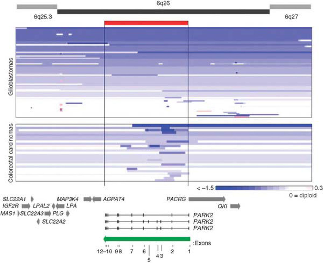
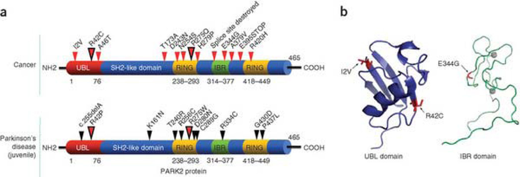
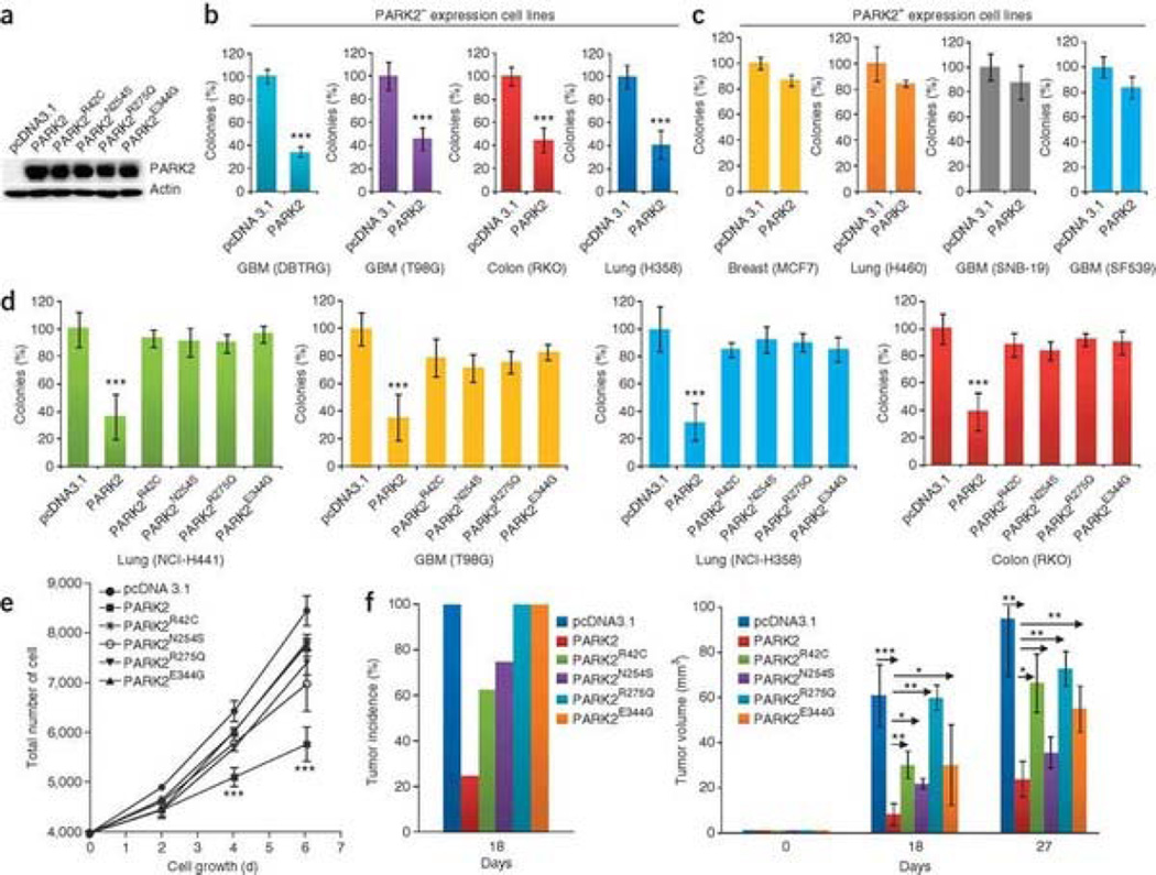
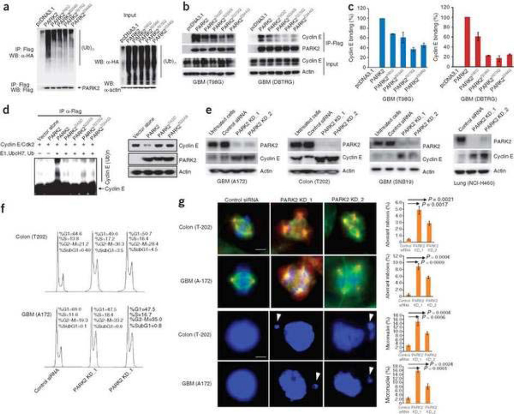
Mutation of the gene PARK2, which encodes an E3 ubiquitin ligase, is the most common cause of early-onset Parkinson's disease. In a search for multisite tumor suppressors, we identified PARK2 as a frequently targeted gene on chromosome 6q25.2-q27 in cancer. Here we describe inactivating somatic mutations and frequent intragenic deletions of PARK2 in human malignancies. The PARK2 mutations in cancer occur in the same domains, and sometimes at the same residues, as the germline mutations causing familial Parkinson's disease. Cancer-specific mutations abrogate the growth-suppressive effects of the PARK2 protein. PARK2 mutations in cancer decrease PARK2's E3 ligase activity, compromising its ability to ubiquitinate cyclin E and resulting in mitotic instability. These data strongly point to PARK2 as a tumor suppressor on 6q25.2-q27. Thus, PARK2, a gene that causes neuronal dysfunction when mutated in the germline, may instead contribute to oncogenesis when altered in non-neuronal somatic cells.


Figures

Figure 1
Figure 1. Diversity of deletions at the PARK2 locus in colon cancer and GBM
Array CGH segmentation map showing GBM (TCGA) and colon cancers (Memorial Sloan-Kettering Cancer Center) for the area surrounding PARK2 on chromosome 6. Analysis and scores were calculated as previously described. Tumors are sorted by amount of loss at the PARK2 locus for convenient viewing. Only tumors showing loss on 6q are shown. The color gradient depicts the extent of copy number loss. The position and boundaries of the PARK2 gene (red bar) are indicated. PARK2 direction and individual exons are labeled (green arrow). Surrounding genes are indicated with gray arrows.
Figure 2
Figure 2. Somatic mutations of PARK2 in human cancers
(a) Summary of PARK2 mutations found in cancer (top) and early-onset Parkinson's disease (bottom). Small arrows show the location of mutations and corresponding amino acid changes. Larger dual-color arrows indicate amino acids that are affected in both cancer and PD; resultant amino acids are different. Mutations cluster in similar regions in both cancer and PD. (b) Structural analysis of cancer-specific mutations in the UBL (left) and IBR (right) domains. Ribbon diagram is shown (with alpha helices and beta sheets). Mutations are shown in red and labeled. Gray circles represent zinc atoms.
Figure 3
Figure 3. Functional analysis of somatic PARK2 mutations in human cancer cell lines
(a) Protein blot showing expression of WT PARK2 and PARK2 with four cancer-specific mutations. Representative data for transfection into T98G are shown. pcDNA3.1, vector-only control. (b) Reconstitution of WT PARK2 suppresses colony-forming ability of human cancer cells lacking PARK2 expression. All assays performed in triplicate. Error bars, ± 1 s.d. ***P < 0.001 (Student's t-test) in all cases. (c) Specificity of PARK2 suppressive effects on colony formation. WT PARK2 was transfected into PARK2-expressing cell lines. Suppressive effects on colony formation are minimal in PARK2+ lines. P > 0.1 (Student's t-test) for all experiments. Error bars, ± 1 s.d. (d) Tumor-derived mutations compromise the colony-forming ability of PARK2 in cancer cells. WT or mutant PARK2 was transfected into the cells indicated. All experiments performed in triplicate. ***P < 0.001 (ANOVA) for all mutants. Error bars, ± 1 s.d. (e) Reconstitution of PARK2 reduces growth rate in cancer cells. DBTRG cells were transfected with each of the constructs shown. All experiments were performed in triplicate. ***P < 0.0001 (ANOVA) for WT PARK2 compared to all others. Error bars, ± 1 s.d. (f) PARK2 reconstitution results in decreased tumor growth in vivo. DBTRG glioma cells stably transfected with vector alone, WT PARK2 and four PARK2 mutants were injected as xenografts. Tumor incidence (left) and tumor size (right) are shown (n= 16). Days on x-axis refer to days following injection of cells into animals. All experiments were performed in duplicate. Arrows indicate comparisons made. *P < 0.05, **P < 0.01, ***P < 0.001 (ANOVA). Error bars, ± 1 s.d.
Figure 4
Figure 4. PARK2 cancer-specific mutations compromise ubiquitination activity
(a) Tumor-derived mutations disrupt PARK2-mediated ubiquitination in cancer cells. T98G cells were transfected with hemagglutinin-ubiquitin (HA-Ub), vector only (pcDNA3.1), WT PARK2 (Flag-tagged) or one of four mutant PARK2 cDNAs (Flag-tagged). Assay was performed as previously described. (b) Cancer-derived mutations of PARK2 decrease association with cyclin E. Indicated cells were treated as above, immunoprecipitated with Flag and detected by protein blot. (c) Quantitation of cyclin E binding efficiency by densitometry. Representative plots shown. For each mutant versus WT, P < 0.05 (Student's t-test). Error bars, ± 1 s.d. (d) Protein blot showing cancer-derived mutations that compromise PARK2-mediated cyclin E ubiquitination in vitro (left). Expression of WT PARK2 but not mutant PARK2 decreases cyclin E levels (right). (e) Knockdown of PARK2 results in increased cyclin E levels. Cells indicated were transfected with PARK2 siRNAs or scrambled siRNA controls and protein blots were performed. (f) Flow cytometry analysis of the indicated cells following PARK2 knockdown. Experiments were performed in triplicate. Representative results are shown. (g) Knockdown of PARK2 results in multipolar spindles and increased frequency of abnormal mitoses (top two rows) and the development of micronuclei (bottom two rows, white arrows). Examples for indicated cells shown using siRNAs targeting PARK2 and scrambled siRNA controls. Red, γ-tubulin; green, α-tubulin. Graphs show quantitation of experiments. Black arrows indicate comparisons made and corresponding P values (Student's t-test). White scale bar for top two rows, 15 µm; bottom two rows, 5 µm. Error bars, ± 1 s.d.

References

    1. Kitada T, et al. Mutations in the parkin gene cause autosomal recessive juvenile parkinsonism. Nature. 1998;392:605–608. - PubMed
    1. Lücking CB, et al. Association between early-onset Parkinson's disease and mutations in the parkin gene. N. Engl J. Med. 2000;342:1560–1567. - PubMed
    1. Fearnley JM, Lees AJ. Ageing and Parkinson's disease: substantia nigra regional selectivity. Brain. 1991;114:2283–2301. - PubMed
    1. Samii A, Nutt JG, Ransom BR. Parkinson's disease. Lancet. 2004;363:1783–1793. - PubMed
    1. Abbas N, et al. A wide variety of mutations in the parkin gene are responsible for autosomal recessive parkinsonism in Europe. French Parkinson's Disease Genetics Study Group and the European Consortium on Genetic Susceptibility in Parkinson's Disease. Hum. Mol. Genet. 1999;8:567–574. - PubMed

Publication types

MeSH terms

Associated data