. 2010 Mar 4;29(9):1270-9.
doi: 10.1038/onc.2009.427. Epub 2009 Nov 30.

Inactivation of HAUSP in vivo modulates p53 function

Affiliations

Inactivation of HAUSP in vivo modulates p53 function

N Kon et al. Oncogene. .

Abstract

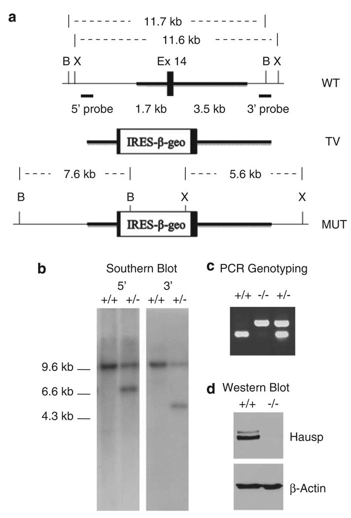
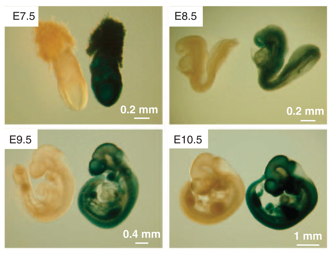
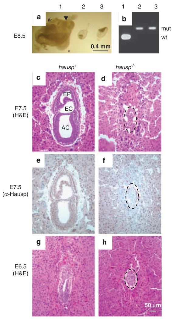
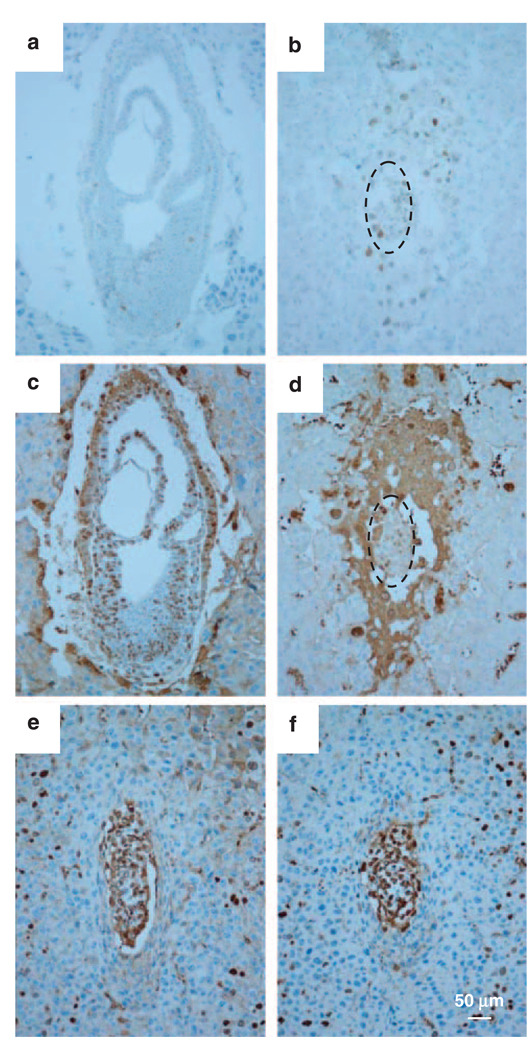
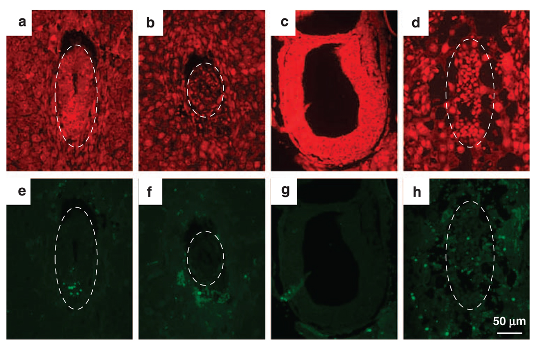
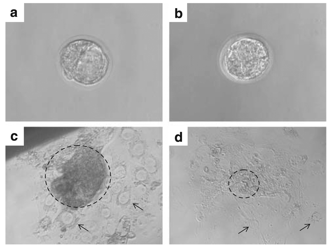
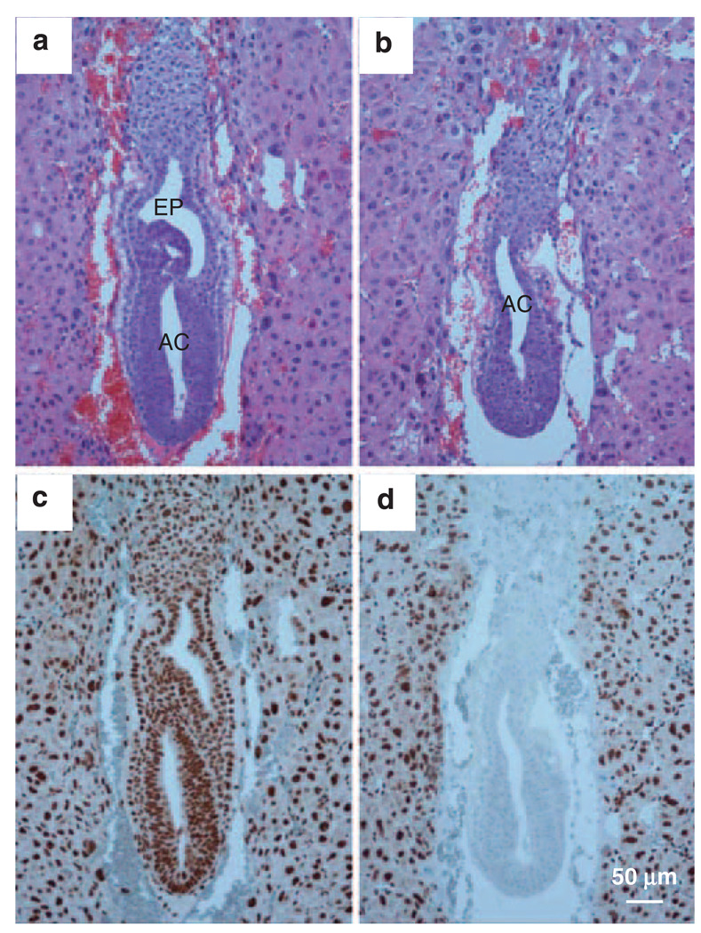
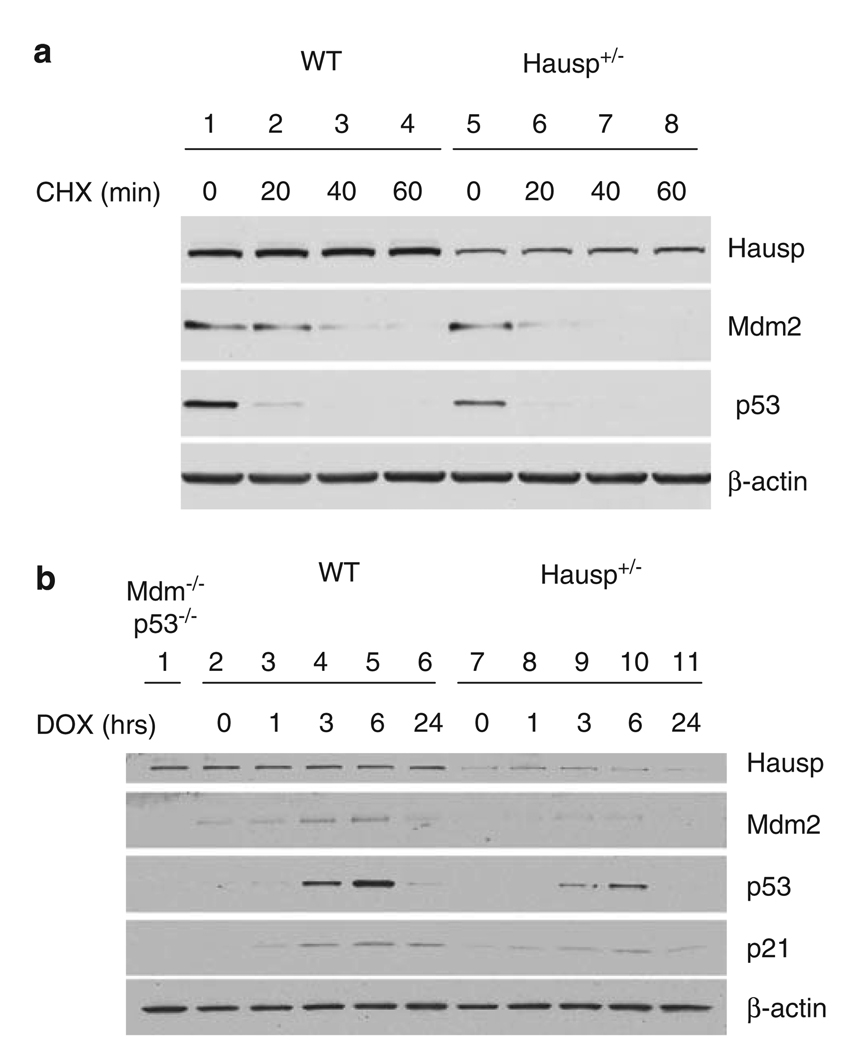
Hausp is a deubiquitinase that has been shown to regulate the p53-Mdm2 pathway. Cotransfection of p53 and Hausp stabilizes p53 through the removal of ubiquitin moieties from polyubiquitinated p53. Interestingly, knockout or RNA interference-mediated knockdown of Hausp in human cells also resulted in the stabilization of p53 due to the destabilization of Mdm2, suggesting a dynamic role of Hausp in p53 activation. To understand the physiological functions of Hausp, we generated hausp knockout mice. Hausp knockout mice die during early embryonic development between embryonic days E6.5 and E7.5. The hausp knockout embryos showed p53 activation, but no apparent increase in apoptosis. Embryonic lethality was caused by a dramatic reduction in proliferation and termination in development, in part due to p53 activation and/or abrogation of p53-independent functions. Although deletion of p53 did not completely rescue the embryonic lethality of the hausp knockout, embryonic development was extended in both hausp and p53 double knockout embryos. These data show that Hausp has a critical role in regulating the p53-Mdm2 pathway.


Conflict of interest statement

Conflict of interest

The authors declare no conflict of interest.

Figures

Figure 1
Figure 1
Targeted disruption of the mouse hausp gene. (a) Diagram of the hausp genomic region containing exon 14 is shown with restriction fragments of BamHI and XbaI indicated. Targeting vector contains IRES-lac Z and a neo cassette was inserted into exon 14, flanking with 1.7 kb 5′-homology and 3.5kb 3′-homology (thicker lines). The diagram of the targeted mutant allele is shown, with altered restriction fragments of BamHI and XbaI after correct gene targeting. (b) Genotyping by Southern blot using genomic DNA digested by BamHI and XbaI, respectively. Hybridization with a 5′-external probe detected an 11.7kb wild-type band and an additional 7.6-kb mutant band in heterozygote mouse. Hybridization using 3′-external probe detected an 11.6 wild-type band and a 5.6-kb mutant band. (c) Genotyping by PCR showed a 202-bp band for wild-type allele and a 433-bp band for mutant allele, simultaneously. (d) The Hausp protein is absent in hausp knockout embryo. Protein extracts prepared from wild-type embryo and hausp knockout embryo at day E8.5, were analyzed by western blot using an anti-Hausp polyclonal antibody and an anti-β-actin monoclonal antibody. The Hausp protein was shown absent in hausp knockout embryo, whereas the total proteins, indicated by β-actin level, were comparable. B, BamHI; X, XbaI.
Figure 2
Figure 2
Expression pattern of Hausp during mouse early embryonic development. The inserted IRES-lacZ allowed the bicistronic expression of β-galactosidase under the control of hausp promoter, which can be detected by staining with X-gal. Embryos from embryonic days E7.5, E8.5, E9.5 and E10.5 were collected and stained with X-gal. As indicated by blue color, Hausp was shown expressed ubiquitously during early embryonic development. Wild-type embryos were used as negative controls for staining.
Figure 3
Figure 3
Phenotypes of hausp knockout embryos. (a) Embryos at day E8.5 from the hausp heterozygote intercross are shown. Embryo no. 1 shows head fold (arrow), somites (*) and tail (arrow head), which are typically present in wild-type embryos at this stage; embryo nos 2 and 3 were abnormal, showing severely reduced cell mass and no advanced structures. (b) Genotyping results for the embryos showed in panel a, embryo no. 1 was wild-type and embryo nos 2 and 3 were hausp knockout embryos. (c) Deciduae from earlier developmental stages were analyzed by histology and immunostaining. Normal embryos at day E7.5 showed development of cavities, such as amniotic cavity (AC), exocoelom (EC) and ectoplacental cavity (EP). (d) Abnormal embryo at day E7.5 failed to develop any noticeable structures. Embryonic cell numbers were greatly reduced (dotted circle). (e) Genotyping of embryos from early embryonic development was performed by immunostaining using a polyclonal antibody anti-Hausp C-terminus. Embryonic cells from the embryo shown in panel c showed brown nuclear staining, suggesting it was either a wild-type or a hausp heterozygote embryo. (f) Embryonic cells (dotted circle) from panel d showed no Hausp staining, suggesting it was a hausp knockout embryo. The surrounding cells from the deciduae were stained by the Hausp antibody, as they were derived from a hasup heterozygote mother. (g) Wild-type embryos at E6.5 show formation of proamniotic cavity, and (h) hausp knockout embryo at E6.5 is smaller and delayed in development. Dotted circles in panels d, f and h, outline the hausp knockout embryos.
Figure 4
Figure 4
Activation of p53 and assay of proliferative activity by BrdU incorporation. Activation of p53 was detected by immunostaining using anti-p53 polyclonal antibody. (a) The wild-type embryos at E7.5 from showed a few p53-positive cells, in contrast, (b) almost all embryonic cells were positive for p53 in hausp knockout embryo. Proliferative activity in embryos was determined by BrdU staining after BrdU incorporation. (c) More than 70% of cells in wild-type embryos at day E7.5 showed BrdU-positive staining, whereas (d) hausp knockout embryos had markedly reduced number of BrdU-positive cells, which was ~35%. However, BrdU-positive cells in wild-type embryo (e) and hausp knockout embryos (f) from day E6.5 were comparable. Dotted circles in panels b and d outline the hausp knockout embryos. BrdU, 5-bromo-2-deoxyuridine.
Figure 5
Figure 5
Detection of apoptotic cells by TdT-mediated dUTP Nick-End Labeling (TUNEL) assay. Embryos from hausp heterozygote intercross were fixed and sectioned. The apoptotic cells were detected by TUNEL, followed by fluorescence microscopy (e–h). Nuclei were counter stained red by propidine iodide (a–d). Wild-type embryos are shown in panels a and e from day E6.5 and in panels c and g from day E7.5. Hausp knockout embryos are shown in panels b and f from day E6.5 and in panels d and h from day E7.5. There were minimum numbers of apoptotic cells detected in both wild-type embryos and hausp knockout embryos. Only wild-type embryo at day E6.5 (e) showed some apoptotic cells, which was likely normal during embryonic development. Dotted circles outline the embryos.
Figure 6
Figure 6
Proliferative activity assay by blastocyst outgrowth in vitro. Blastocysts, (a) wild-type and (b) hausp knockout embryos, were collected from hausp heterozygote intercross at day E3.5 and cultured individually in vitro for 5 days. (c) Wild-type blastocysts showed prominent outgrowth of the embryonic cells (dotted circle) from the inner cell mass of the blastocyst on day 5. Trophoblastic giant cells are indicated by arrows. (d) Hausp knockout embryo showed virtually no outgrowth of the embryonic cells (dotted circle), the majority of the surviving cells were trophoblastic giant cells (arrows).
Figure 7
Figure 7
Deletion of p53 extended the embryonic development in hausp knockout embryo. Embryos from the intercross of hausp heterozygote, p53 homozygote mice were collected at day E7.5. The genotype of embryos shown in (a) and (b) was determined by immunostaining using anti-Hausp C-terminus antibody shown in (c) and (d), respectively. The hausp-positive, p53 knockout embryos (panel a) showed normal development, such as formation of amniotic cavity (AC) and ectoplacental cavity (EP). Similarly, the hausp and p53 double knockout embryo (b) also showed formation of amniotic cavity. However, the development of ectoplacental cavity was missing or delayed.
Figure 8
Figure 8
Stability of Mdm2 and p53 in hausp heterozygote MEF cells. (a) Comparison of the protein half-lives of p53 and mdm2 in MEF cells. Total cell extracts were made from wild-type MEF cells (lanes 1–4) and hausp heterozygote MEF cells (lanes 5–8) after cells were incubated for 0 (lanes 1 and 5), 20 (lanes 2 and 6), 40 (lanes 3 and 7) and 60 min (lanes 4 and 8) in the presence of 10 µg/ml cycloheximide. The protein levels of Hausp, p53 and Mdm2, were determined by western blot and sensitive enhanced chemiluminesence (ECL). The level of β-actin was used as internal control for total protein. (b) Reduced activation of p53 in hausp heterozygote MEF cells after DNA damage. Total cell extracts were made from wild-type MEF cells (lanes 2–6) and hausp heterozygote MEF cells (lanes 7–11) after cells were incubated for 1 h (lanes 3 and 8), 3 h (lanes 4 and 9), 6 h (lanes 6 and 10) and 24 h (lanes 7 and 11) in the presence of 0.2 µg/ml Doxorubicin, respectively. Cell extracts from wild-type and hausp heterozygote MEF cells without doxorubicin treatment were loaded in lanes 2 and 7, respectively. Cell extracts from p53 and mdm2 double knockout MEF cells were used to facilitate identification of p53 and Mdm2 (lane 1). The protein levels of Hausp, p53, Mdm2 and p53 downstream target p21 were determined by western blot and regular ECL. The level of β-actin was used as internal control for total proteins. MEF, mouse embryonic fibroblast.

References

    1. Amerik AY, Li S, Hochstrasser M. Analysis of the deubiquitinating enzymes of the yeast Saccharomyces cerevisiae. Biol Chem. 2000;381:981–992. - PubMed
    1. Babu JR, Jeganathan KB, Baker DJ, Wu X, Kang-Decker N, van Deursen JM. Rae1 is an essential mitotic checkpoint regulator that cooperates with Bub3 to prevent chromosome missegregation. J Cell Biol. 2003;3:341–353. - PMC - PubMed
    1. Becker K, Marchenko ND, Palacios G, Moll UM. A role of HAUSP in tumor suppression in a human colon carcinoma xenograft model. Cell Cycle. 2008;7:1205–1213. - PMC - PubMed
    1. Chen D, Kon N, Li M, Zhang W, Qin J, Gu W. ARF-BP1/ Mule is a critical mediator of the ARF tumor suppressor. Cell. 2005;121:1071–1083. - PubMed
    1. Chen X, Ko LJ, Jayaraman L, Prives C. p53 levels, functional domains, and DNA damage determine the extent of the apoptotic response of tumor cells. Genes Dev. 1996;1:2438–2451. - PubMed

Publication types

MeSH terms