Akt promotes endocardial-mesenchyme transition
- PMID: 19946410
- PMCID: PMC2776235
- DOI: 10.1186/2040-2384-1-2
Akt promotes endocardial-mesenchyme transition
Abstract
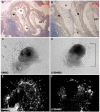
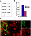
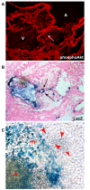
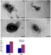
Endothelial to mesenchyme transition (EndMT) can be observed during the formation of endocardial cushions from the endocardium, the endothelial lining of the atrioventricular canal (AVC), of the developing heart at embryonic day 9.5 (E9.5). Many regulators of the process have been identified; however, the mechanisms driving the initial commitment decision of endothelial cells to EndMT have been difficult to separate from processes required for mesenchymal proliferation and migration. We have several lines of evidence that suggest a central role for Akt signaling in committing endothelial cells to enter EndMT. Akt1 mRNA was restricted to the endocardium of endocardial cushions while they were forming. The PI3K/Akt signaling pathway is necessary for mesenchyme outgrowth, as sprouting was inhibited in AVC explant cultures treated with the PI3K inhibitor LY294002. Furthermore, endothelial marker, VE-cadherin, was downregulated and mesenchyme markers, N-cadherin and Snail, were induced in response to expression of a constitutively active form of Akt1 (myrAkt1) in endothelial cells. Finally, we isolated the function of Akt1 signaling in the commitment to the transition using a transgenic model where myrAkt1 was pulsed only in endocardial cells and turned off after EndMT initiation. In this way, we determined that increased Akt signaling in the endocardium drives EndMT and discounted its other functions in cushion mesenchymal cells.
Figures


Similar articles
-
Impairment of endothelial-mesenchymal transformation during atrioventricular cushion formation in Tmem100 null embryos.Dev Dyn. 2015 Jan;244(1):31-42. doi: 10.1002/dvdy.24216. Epub 2014 Nov 7. Dev Dyn. 2015. PMID: 25318679
-
Endothelial expression of bone morphogenetic protein receptor type 1a is required for atrioventricular valve formation.Ann Thorac Surg. 2008 Jun;85(6):2090-8. doi: 10.1016/j.athoracsur.2008.02.027. Ann Thorac Surg. 2008. PMID: 18498827
-
Mechanical strain triggers endothelial-to-mesenchymal transition of the endocardium in the immature heart.Pediatr Res. 2022 Sep;92(3):721-728. doi: 10.1038/s41390-021-01843-6. Epub 2021 Nov 26. Pediatr Res. 2022. PMID: 34837068 Free PMC article.
-
Epithelial-mesenchymal transformations in early avian heart development.Acta Anat (Basel). 1996;156(3):173-86. doi: 10.1159/000147845. Acta Anat (Basel). 1996. PMID: 9124035 Review.
-
Molecular regulation of atrioventricular valvuloseptal morphogenesis.Circ Res. 1995 Jul;77(1):1-6. doi: 10.1161/01.res.77.1.1. Circ Res. 1995. PMID: 7788867 Review.
Cited by
-
ErbB signaling in cardiac development and disease.Semin Cell Dev Biol. 2010 Dec;21(9):929-35. doi: 10.1016/j.semcdb.2010.09.011. Epub 2010 Oct 7. Semin Cell Dev Biol. 2010. PMID: 20933094 Free PMC article. Review.
-
Endothelial to Mesenchymal Transition in Cardiovascular Disease: JACC State-of-the-Art Review.J Am Coll Cardiol. 2019 Jan 22;73(2):190-209. doi: 10.1016/j.jacc.2018.09.089. J Am Coll Cardiol. 2019. PMID: 30654892 Free PMC article. Review.
-
Inflammatory cytokines promote mesenchymal transformation in embryonic and adult valve endothelial cells.Arterioscler Thromb Vasc Biol. 2013 Jan;33(1):121-30. doi: 10.1161/ATVBAHA.112.300504. Epub 2012 Oct 25. Arterioscler Thromb Vasc Biol. 2013. PMID: 23104848 Free PMC article.
-
The polycomb group protein EZH2 induces epithelial-mesenchymal transition and pluripotent phenotype of gastric cancer cells by binding to PTEN promoter.J Hematol Oncol. 2018 Jan 15;11(1):9. doi: 10.1186/s13045-017-0547-3. J Hematol Oncol. 2018. PMID: 29335012 Free PMC article.
-
Lysyl oxidase-like 2 is a regulator of angiogenesis through modulation of endothelial-to-mesenchymal transition.J Cell Physiol. 2019 Jul;234(7):10260-10269. doi: 10.1002/jcp.27695. Epub 2018 Nov 1. J Cell Physiol. 2019. PMID: 30387148 Free PMC article.
References
-
- Nakajima Y, Yamagishi T, Hokari S, Nakamura H. Mechanisms involved in valvuloseptal endocardial cushion formation in early cardiogenesis: roles of transforming growth factor (TGF)-beta and bone morphogenetic protein (BMP) Anat Rec. 2000;258:119–127. doi: 10.1002/(SICI)1097-0185(20000201)258:2<119::AID-AR1>3.0.CO;2-U. - DOI - PubMed
LinkOut - more resources
Full Text Sources
Research Materials
Miscellaneous

