

Evidence for the progression through S-phase in the ectopic cell cycle re-entry of neurons in Alzheimer disease
- PMID: 19946466
- PMCID: PMC2783633
- DOI: 10.18632/aging.100044
Evidence for the progression through S-phase in the ectopic cell cycle re-entry of neurons in Alzheimer disease
Abstract
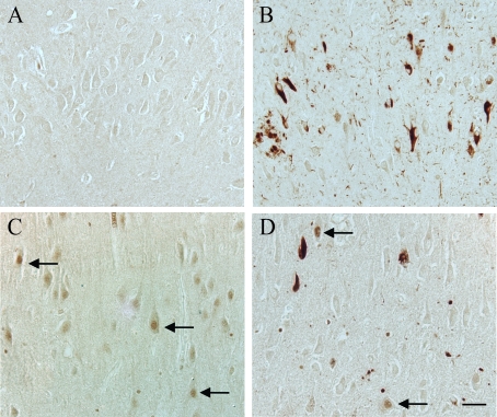
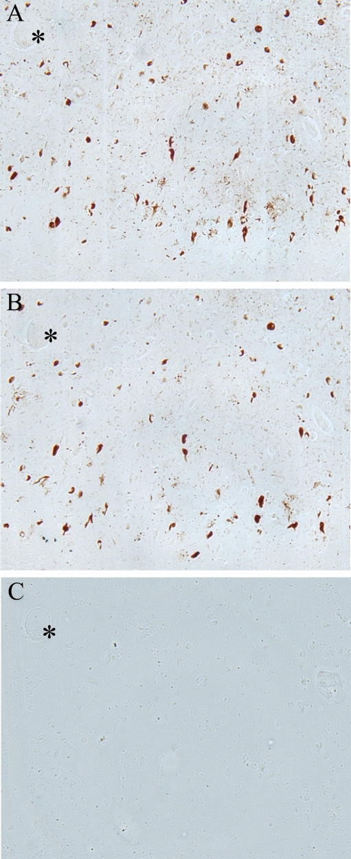
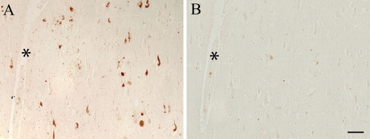
Aberrant neuronal re-entry into the cell cycle is emerging as a potential pathological mechanism in Alzheimer disease (AD). However, while cyclins, cyclin dependent kinases (CDKs), and other mitotic factors are ectopically expressed in neurons, many of these proteins are also involved in other pathological and physiological processes, generating continued debate on whether such markers are truly indicative of a bona fide cell cycle process. To address this issue, here we analyzed one of the minichromosome maintenance (Mcm) proteins that plays a role in DNA replication and becomes phosphorylated by the S-phase promoting CDKs and Cdc7 during DNA synthesis. We found phosphorylated Mcm2 (pMcm2) markedly associated with neurofibrillary tangles, neuropil threads, and dystrophic neurites in AD but not in aged-matched controls. These data not only provide further evidence for cell cycle aberrations in AD, but the cytoplasmic, rather than nuclear, localization of pMcm2 suggests an abnormal cellular distribution of this important replication factor in AD that may explain resultant cell cycle stasis and consequent neuronal degeneration.
Keywords: Alzheimer disease; DNA replication; cell cycle; minichromosome maintenance protein; neurodegeneration.
Conflict of interest statement
Dr. Smith is, or has in the past been, a paid consultant for, owns equity or stock options in and/or receives grant funding from Canopus BioPharma, Medivation, Neurotez, Neuropharm, Panacea Pharmaceuticals, and Voyager Pharmaceuticals. Dr. Perry is, or has in the past been, a paid consultant for and/or owns equity or stock options in Takeda Pharmaceuticals, Voyager Pharmaceuticals, Panacea Pharmaceuticals and Neurotez Pharmaceuticals.
Figures

References
-
- Smith MA. Alzheimer disease. Int Rev Neurobiol. 1998;42:1–54. - PubMed
-
- Folstein MF, Bylsma FW. In: Alzheimer disease. Terry RD, Katzman R, Bick KL, Sisodia SS, editors. Philadelphia: Lippincott Williams & Wilkins; 1999. Noncognitive symptoms of Alzheimer disease ; pp. 25–37.
-
- Morris JC. In: Alzheimer disease. Terry RD, Katzman R, Bick KL, Sisodia SS, editors. Philadelphia: Lippincott Williams & Wilkins; 1999. Clinical presentation and course of Alzheimer disease ; pp. 11–24.
-
- West MJ, Kawas CH, Stewart WF, Rudow GL, Troncoso JC. Hippocampal neurons in pre-clinical Alzheimer's disease. Neurobiol Aging. 2004;25:1205–1212. - PubMed
-
- Zhu X, Lee HG, Perry G, Smith MA. Alzheimer disease, the two-hit hypothesis: an update. Biochim Biophys Acta. 2007;1772:494–502. - PubMed
Publication types
MeSH terms
Substances
Grants and funding
LinkOut - more resources
Full Text Sources
Other Literature Sources
Medical
Miscellaneous