

Alterations in gene expression and sensitivity to genotoxic stress following HdmX or Hdm2 knockdown in human tumor cells harboring wild-type p53
- PMID: 19946469
- PMCID: PMC2783638
- DOI: 10.18632/aging.100008
Alterations in gene expression and sensitivity to genotoxic stress following HdmX or Hdm2 knockdown in human tumor cells harboring wild-type p53
Abstract
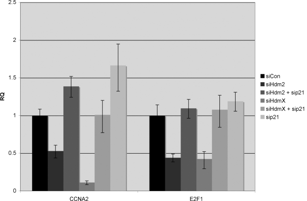
While half of all human tumors possess p53 mutations, inactivation of wild-type p53 can also occur through a variety of mechanisms that do not involve p53 gene mutation or deletion. Our laboratory has been interested in tumor cells possessing wild-type p53 protein and elevated levels of HdmX and/or Hdm2, two critical negative regulators of p53 function. In this study we utilized RNAi to knockdown HdmX or Hdm2 in MCF7 human breast cancer cells, which harbor wild-type p53 and elevated levels of HdmX and Hdm2 then examined gene expression changes and effects on cell growth.Cell cycle and growth assays confirmed that the loss of either HdmX or Hdm2 led to a significant growth inhibition and G1cell cycle arrest. Although the removal of overexpressed HdmX/2 appears limited to an anti-proliferative effect in MCF7cells, the loss of HdmX and/or Hdm2 enhanced cytotoxicity in these same cells exposed to DNA damage. Through the use of Affymetrix GeneChips and subsequent RT-qPCR validations, we uncovered a subset of anti-proliferative p53 target genes activated upon HdmX/2 knockdown. Interestingly, a second set of genes, normally transactivated by E2F1 as cells transverse the G1-S phase boundary, were found repressed in a p21-dependent manner following HdmX/2 knockdown.Taken together, these results provide novel insights into the reactivation of p53 in cells overexpressing HdmX and Hdm2.
Keywords: Hdm2; HdmX; RNAi; gene expression profiling; p53.
Conflict of interest statement
The authors of this manuscript have no conflict of interests to declare.
Figures


Similar articles
-
HdmX overexpression inhibits oncogene induced cellular senescence.Cell Cycle. 2010 Aug 15;9(16):3376-82. doi: 10.4161/cc.9.16.12779. Epub 2010 Aug 23. Cell Cycle. 2010. PMID: 20724842 Free PMC article.
-
HDMX-L is expressed from a functional p53-responsive promoter in the first intron of the HDMX gene and participates in an autoregulatory feedback loop to control p53 activity.J Biol Chem. 2010 Sep 17;285(38):29111-27. doi: 10.1074/jbc.M110.129726. Epub 2010 Jul 20. J Biol Chem. 2010. PMID: 20659896 Free PMC article.
-
Levels of HdmX expression dictate the sensitivity of normal and transformed cells to Nutlin-3.Cancer Res. 2006 Mar 15;66(6):3169-76. doi: 10.1158/0008-5472.CAN-05-3832. Cancer Res. 2006. PMID: 16540668
-
HDM2 and HDMX Proteins in Human Cancer.Klin Onkol. 2018 Winter;31(Suppl 2):63-70. doi: 10.14735/amko20182S63. Klin Onkol. 2018. PMID: 31023026 Review. English.
-
Posttranscriptional regulation of p53 and its targets by RNA-binding proteins.Curr Mol Med. 2008 Dec;8(8):845-9. doi: 10.2174/156652408786733748. Curr Mol Med. 2008. PMID: 19075680 Free PMC article. Review.
Cited by
-
Combined study on clastogenic, aneugenic and apoptotic properties of doxorubicin in human cells in vitro.J Biol Res (Thessalon). 2018 Oct 11;25:17. doi: 10.1186/s40709-018-0089-z. eCollection 2018 Dec. J Biol Res (Thessalon). 2018. PMID: 30338246 Free PMC article.
-
Recent progress in targeting cancer.Aging (Albany NY). 2011 Dec;3(12):1154-62. doi: 10.18632/aging.100421. Aging (Albany NY). 2011. PMID: 22228887 Free PMC article. Review.
-
Proposed megakaryocytic regulon of p53: the genes engaged to control cell cycle and apoptosis during megakaryocytic differentiation.Physiol Genomics. 2012 Jun 15;44(12):638-50. doi: 10.1152/physiolgenomics.00028.2012. Epub 2012 May 1. Physiol Genomics. 2012. PMID: 22548738 Free PMC article.
-
Mdm2 and MdmX as Regulators of Gene Expression.Genes Cancer. 2012 Mar;3(3-4):264-73. doi: 10.1177/1947601912455331. Genes Cancer. 2012. PMID: 23150759 Free PMC article.
-
HdmX overexpression inhibits oncogene induced cellular senescence.Cell Cycle. 2010 Aug 15;9(16):3376-82. doi: 10.4161/cc.9.16.12779. Epub 2010 Aug 23. Cell Cycle. 2010. PMID: 20724842 Free PMC article.
References
-
- Kubbutat MH, Vousden KH. Keeping an old friend under control: regulation of p53 stability. Mol Med Today. 1998;4:250–256. - PubMed
-
- Vousden KH, X Lu. Live or let die: the cell's response to p53. Nat Rev Cancer. 2002;2:594–604. - PubMed
-
- Marine JC, Jochemsen AG. Mdmx and Mdm2: brothers in arms. Cell Cycle. 2004;3:900–904. - PubMed
-
- Oliner JD, Peitenol JA, Thiagalingam S, Gyuris J, Kinzler KW, Vogelstein B. Oncoprotein MDM2 conceals the activation domain of tumour suppressor p53. Nature. 1993;362:857–860. - PubMed
Publication types
MeSH terms
Substances
Grants and funding
LinkOut - more resources
Full Text Sources
Research Materials
Miscellaneous