Effects of Culturing on the Stability of the Putative Murine Adipose Derived Stem Cells Markers
- PMID: 19946473
- PMCID: PMC2783658
- DOI: 10.2174/1876893800901010054
Effects of Culturing on the Stability of the Putative Murine Adipose Derived Stem Cells Markers
Abstract
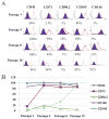
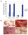
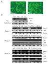
Mesenchymal stem cells have generated much interest because of their potential use in regenerative medicine. The major draw back in the application of these cells is that there is no single marker or markers that have been established to identify and aid in isolating the cells from a variety of other cell types. The commonly expressed mesenchymal stem cell surface antigens include CD44, CD73, CD90.2, CD105, and CD146. In the present study we examined the stability of these surface antigens in culture and their potential application in identifying and isolating murine derived adipose derived stem cells. The data showed that the expression of these markers increased with culturing and appeared to stabilize by passage 8; the cells were sorted positively for the surface markers at this passage. Each subset was maintained in culture and evaluated for differentiation toward osteogenic lineage in vitro and in vivo. The CD73 and CD105 positive cell subsets demonstrated robust differentiation toward osteogenic lineage in vitro; the CD90.2+ cell subset exhibited the least differentiation toward osteogenic lineage. Assessment of the cell subpopulations for in vivo differentiation demonstrated that all the cell subsets exhibited potential to differentiate into osteoblasts. Taken together, these data suggest that this panel of markers although useful in identifying cells with potential to differentiate toward osteogenic lineage, cannot prospectively be used for enriching for ADSC from a variety of other cell types.
Figures


Similar articles
-
Discrete adipose-derived stem cell subpopulations may display differential functionality after in vitro expansion despite convergence to a common phenotype distribution.Stem Cell Res Ther. 2016 Dec 1;7(1):177. doi: 10.1186/s13287-016-0435-8. Stem Cell Res Ther. 2016. PMID: 27906060 Free PMC article.
-
Characterization of mesenchymal stem cell subpopulations from human amniotic membrane with dissimilar osteoblastic potential.Stem Cells Dev. 2013 Apr 15;22(8):1275-87. doi: 10.1089/scd.2012.0359. Epub 2013 Jan 29. Stem Cells Dev. 2013. PMID: 23211052
-
Electromagnetic Fields Generated by the IteraCoil Device Differentiate Mesenchymal Stem Progenitor Cells Into the Osteogenic Lineage.Bioelectromagnetics. 2022 May;43(4):245-256. doi: 10.1002/bem.22401. Epub 2022 Apr 7. Bioelectromagnetics. 2022. PMID: 35391494 Free PMC article.
-
Cell Surface Markers on Adipose-Derived Stem Cells: A Systematic Review.Curr Stem Cell Res Ther. 2017;12(6):484-492. doi: 10.2174/1574888X11666160429122133. Curr Stem Cell Res Ther. 2017. PMID: 27133085
-
Osteogenic Potential of Mouse Adipose-Derived Stem Cells Sorted for CD90 and CD105 In Vitro.Stem Cells Int. 2014;2014:576358. doi: 10.1155/2014/576358. Epub 2014 Sep 15. Stem Cells Int. 2014. PMID: 25302065 Free PMC article.
Cited by
-
Functional Regulatory Mechanisms Underlying Bone Marrow Mesenchymal Stem Cell Senescence During Cell Passages.Cell Biochem Biophys. 2021 Jun;79(2):321-336. doi: 10.1007/s12013-021-00969-y. Epub 2021 Feb 9. Cell Biochem Biophys. 2021. PMID: 33559812
-
Cardiac mesenchymal stem cells contribute to scar formation after myocardial infarction.Cardiovasc Res. 2011 Jul 1;91(1):99-107. doi: 10.1093/cvr/cvr061. Epub 2011 Feb 28. Cardiovasc Res. 2011. PMID: 21357194 Free PMC article.
-
IGF1 Promotes Adipogenesis by a Lineage Bias of Endogenous Adipose Stem/Progenitor Cells.Stem Cells. 2015 Aug;33(8):2483-95. doi: 10.1002/stem.2052. Epub 2015 May 27. Stem Cells. 2015. PMID: 26010009 Free PMC article.
-
Unravelling the retention of proliferation and differentiation potency in extensive culture of human subcutaneous fat-derived mesenchymal stem cells in different media.Cell Prolif. 2012 Dec;45(6):516-26. doi: 10.1111/j.1365-2184.2012.00843.x. Cell Prolif. 2012. PMID: 23106299 Free PMC article.
-
Isolation and Characterization of Mouse Adipose-Derived Stromal/Stem Cells.Methods Mol Biol. 2025;2938:119-123. doi: 10.1007/978-1-0716-4607-6_14. Methods Mol Biol. 2025. PMID: 40445287
References
-
- Arthur HM, Ure J, Smith AJ, et al. Endoglin, an ancillary TGFbeta receptor, is required for extraembryonic angiogenesis and plays a key role in heart development. Dev Biol. 2000;217(1):42–53. - PubMed
-
- Krupinski J, Turu MM, Luque A, Badimon L, Slevin M. Increased PrPC expression correlates with endoglin (CD105) positive microvessels in advanced carotid lesions. Acta Neuropathol. 2008;116(5):537–45. - PubMed
-
- Pérez-Gómez E, Eleno N, López-Novoa JM, et al. Characterization of murine S-endoglin isoform and its effects on tumor development. Oncogene. 2005;24(27):4450–61. - PubMed
-
- Shih IM. The role of CD146 (Mel-CAM) in biology and pathology. J Pathol. 1999;189(1):4–11. - PubMed
Grants and funding
LinkOut - more resources
Full Text Sources
Research Materials
Miscellaneous
