

An atypical case of hepatic cavernous hemangioma
- PMID: 19946491
- PMCID: PMC2783136
- DOI: 10.1186/1757-1626-2-181
An atypical case of hepatic cavernous hemangioma
Abstract
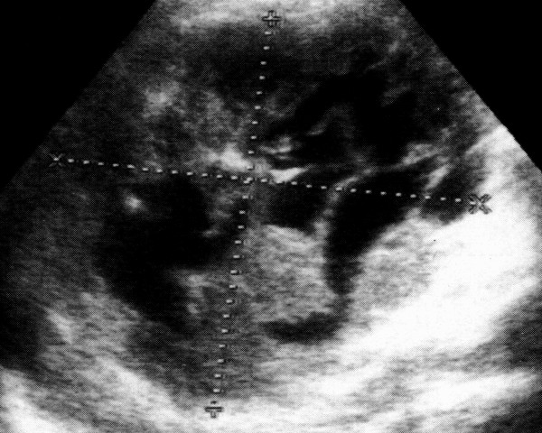
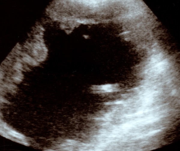
Introduction: The case of an atypical hepatic angiocavernoma is referred. The lesion, first described as a hypoechogenic area compared to the surrounding parenchyma, with anechogenic shoots inside, suggestive for vascular structures developed one year later into a totally asonic area with frayed margins. This change is very unusual and uncommon for this kind of lesions.
Case presentation: The case of a 74-year old caucasian male, complaining of slight dyspeptic symptoms (post-prandial fullness and bloating) is referred. The routine blood tests were all normal. Abdominal ultrasound showed a large, roughly round-shaped lesion (diameter 14 cm) in the VIII hepatic segment diagnosed as hepatic angiocavernoma, which turned unexpectedly in a cystic like lesion one year later.
Conclusion: The atypical angioma's degeneration could account for one of the causes of the patient's exitus. It could be related to blood seizure by the large hepatic angioma due to the intratumoural haemorrhage.
Figures
Similar articles
-
[Two cases of huge hepatic hemangioma].Fukuoka Igaku Zasshi. 1993 Mar;84(3):91-6. Fukuoka Igaku Zasshi. 1993. PMID: 8477925 Review. Japanese.
-
Endoscopic Ultrasound-Guided Ethanol Injection Associated with Trans-arterial Embolization of a Giant Intra-abdominal Cavernous Hemangioma: Case Report and New Therapeutic Option.J Gastrointest Cancer. 2021 Mar;52(1):381-385. doi: 10.1007/s12029-020-00568-9. Epub 2021 Jan 7. J Gastrointest Cancer. 2021. PMID: 33411258
-
A hepatic sclerosed hemangioma with drastic changes in contrast-enhanced ultrasonography.Clin J Gastroenterol. 2020 Dec;13(6):1252-1257. doi: 10.1007/s12328-020-01194-5. Epub 2020 Jul 23. Clin J Gastroenterol. 2020. PMID: 32705537
-
Adrenal cavernous hemangioma: is presurgical diagnosis with imaging test possible.Arch Esp Urol. 2013 Apr;66(3):313-6. Arch Esp Urol. 2013. PMID: 23648753 English, Spanish.
-
Spontaneous intracapsular hemorrhage of a giant hepatic cavernous hemangioma: a rare case report and literature review.BMC Gastroenterol. 2021 Feb 23;21(1):84. doi: 10.1186/s12876-021-01666-z. BMC Gastroenterol. 2021. PMID: 33622256 Free PMC article. Review.
Cited by
-
Rim 18F-fluorodeoxyglucose uptake of hepatic cavernous hemangioma on positron emission tomography/computed tomography: A case report.World J Clin Cases. 2024 May 6;12(13):2243-2247. doi: 10.12998/wjcc.v12.i13.2243. World J Clin Cases. 2024. PMID: 38808338 Free PMC article.
-
Morphological and dynamic evaluation of complex cystic focal liver lesions by contrast-enhanced ultrasound: current state of the art.J Ultrasound. 2019 Sep;22(3):251-259. doi: 10.1007/s40477-019-00385-2. Epub 2019 May 13. J Ultrasound. 2019. PMID: 31087277 Free PMC article. Review.
-
Cavernous haemangioma with cyst formation coexisting with giant non-parasitic cyst in the liver.BMJ Case Rep. 2022 Sep 13;15(9):e251242. doi: 10.1136/bcr-2022-251242. BMJ Case Rep. 2022. PMID: 36100288 Free PMC article. No abstract available.
References
-
- Brogna A. Ecografia addominale integrata. Lombardo Editore. 2004.
-
- Mungovan JA, Cranon JJ, Vacarro J. Hepatic cavernous hemangiomas: lack of enlargement over time. Radiology. 1994;191:111–113. - PubMed
-
- Okano H, Shiraki K, Inoue H, Ito T, Yamanaka T, Deguchi M, Sugimoto K, Sakai T, Ohmori S, Murata K, Takase K, Nakano T. Natural course of cavernous hepatic hemangioma. Oncol Rep. 2001;8(2):411–4. - PubMed
-
- Rumack Carol M, Wilson Stephanie R, William Charboneau J. Diagnostic Ultrasound. Mosby. 1998.
-
- Moody AR, Wilson SR. Atypical hemangioma: a suggestive sonographic morphology. Radiology. 1993;188:413–417. - PubMed
LinkOut - more resources
Full Text Sources
