

Russell bodies in a skin biopsy: a case report
- PMID: 19946586
- PMCID: PMC2783049
- DOI: 10.1186/1752-1947-3-108
Russell bodies in a skin biopsy: a case report
Abstract
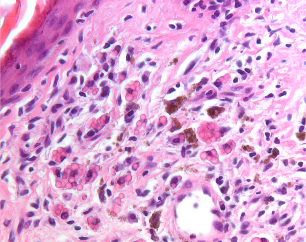
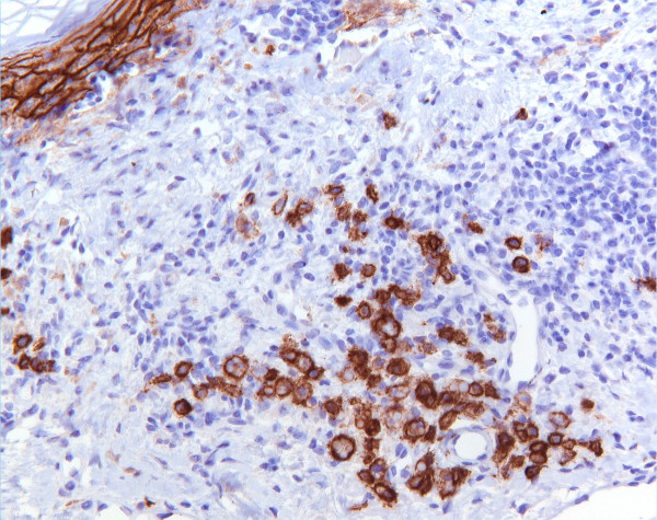
Introduction: The presence of eosinophilic bodies in a skin biopsy can be found in a variety of situations and this may present a challenge to the pathologist. The differential diagnosis of these eosinophilic structures include microorganisms such as histoplasmosis or cryptococcosis, fungi, Michaelis-Gutmann bodies, deposits of amyloid or immunoglobulins, colloid bodies or elastic bodies.
Case presentation: During a routine examination of a skin biopsy with actinic keratosis taken from the cheek of a 61-year-old man, clusters of eosinophilic bodies were seen within an inflammatory infiltrate in the dermis, both intracytoplasmic and extracellular. Using additional immunohistochemical staining, these structures were identified as polyclonal Russell bodies.
Conclusion: The differential diagnosis of intracytoplasmic eosinophilic structures in a skin biopsy includes Russell bodies, an uncommon finding that may be associated with chronic inflammatory conditions.
Figures
Similar articles
-
Prominent dermal accumulation of Russell bodies underlying pseudocarcinomatous hyperplasia with fungal infection.Nagoya J Med Sci. 2023 Feb;85(1):123-126. doi: 10.18999/nagjms.85.1.123. Nagoya J Med Sci. 2023. PMID: 36923611 Free PMC article.
-
Russell body cervicitis: A case report and literature review highlighting diagnostic pitfalls.Case Rep Womens Health. 2025 Apr 15;46:e00707. doi: 10.1016/j.crwh.2025.e00707. eCollection 2025 Jun. Case Rep Womens Health. 2025. PMID: 40297197 Free PMC article.
-
Russell body gastroenteritis: an aberrant manifestation of chronic inflammation in gastrointestinal mucosa.Case Rep Med. 2013;2013:797264. doi: 10.1155/2013/797264. Epub 2013 Oct 3. Case Rep Med. 2013. PMID: 24198839 Free PMC article.
-
Role of In Vivo Reflectance Confocal Microscopy in the Analysis of Melanocytic Lesions.Acta Dermatovenerol Croat. 2018 Apr;26(1):64-67. Acta Dermatovenerol Croat. 2018. PMID: 29782304 Review.
-
Russell Bodies and Russell Body Inflammatory Polyp in the Colorectum: A Review of Clinicopathologic Features.Biomed Res Int. 2018 Jul 29;2018:2845291. doi: 10.1155/2018/2845291. eCollection 2018. Biomed Res Int. 2018. PMID: 30151376 Free PMC article. Review.
Cited by
-
Prominent dermal accumulation of Russell bodies underlying pseudocarcinomatous hyperplasia with fungal infection.Nagoya J Med Sci. 2023 Feb;85(1):123-126. doi: 10.18999/nagjms.85.1.123. Nagoya J Med Sci. 2023. PMID: 36923611 Free PMC article.
-
Localized accumulation of kappa restricted Russell body-containing plasma cells in tonsil.Clin Case Rep. 2019 Aug 9;7(9):1766-1768. doi: 10.1002/ccr3.2372. eCollection 2019 Sep. Clin Case Rep. 2019. PMID: 31534745 Free PMC article.
-
Russell body cervicitis: A case report and literature review highlighting diagnostic pitfalls.Case Rep Womens Health. 2025 Apr 15;46:e00707. doi: 10.1016/j.crwh.2025.e00707. eCollection 2025 Jun. Case Rep Womens Health. 2025. PMID: 40297197 Free PMC article.
References
-
- Weedon D. Skin Pathology. 2. Edinburgh: Churchill Livingstone; 2002. p. 438.
LinkOut - more resources
Full Text Sources
