

cGMP-phosphodiesterase 6, transducin and Wnt5a/Frizzled-2-signaling control cGMP and Ca(2+) homeostasis in melanoma cells
- PMID: 19946729
- PMCID: PMC11115744
- DOI: 10.1007/s00018-009-0214-0
cGMP-phosphodiesterase 6, transducin and Wnt5a/Frizzled-2-signaling control cGMP and Ca(2+) homeostasis in melanoma cells
Retraction in
-
Retraction Note to: cGMP-phosphodiesterase 6, transducin and Wnt5a/Frizzled-2-signaling control cGMP and Ca2+ homeostasis in melanoma cells.Cell Mol Life Sci. 2020 Mar;77(5):963. doi: 10.1007/s00018-019-03437-2. Cell Mol Life Sci. 2020. PMID: 31919573 Free PMC article.
Abstract
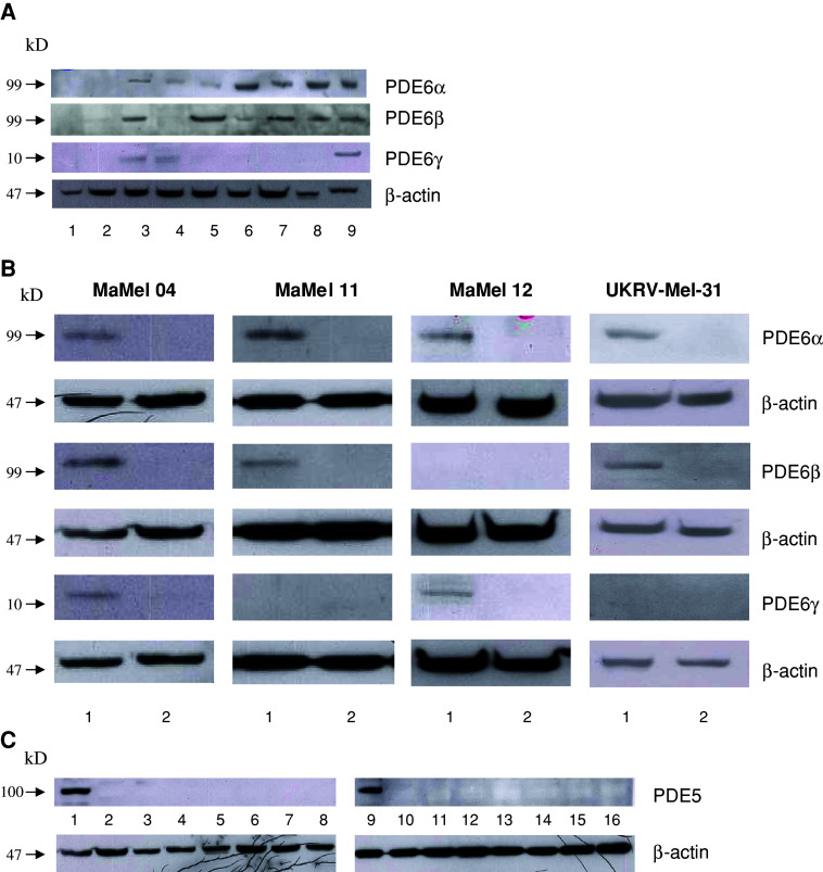
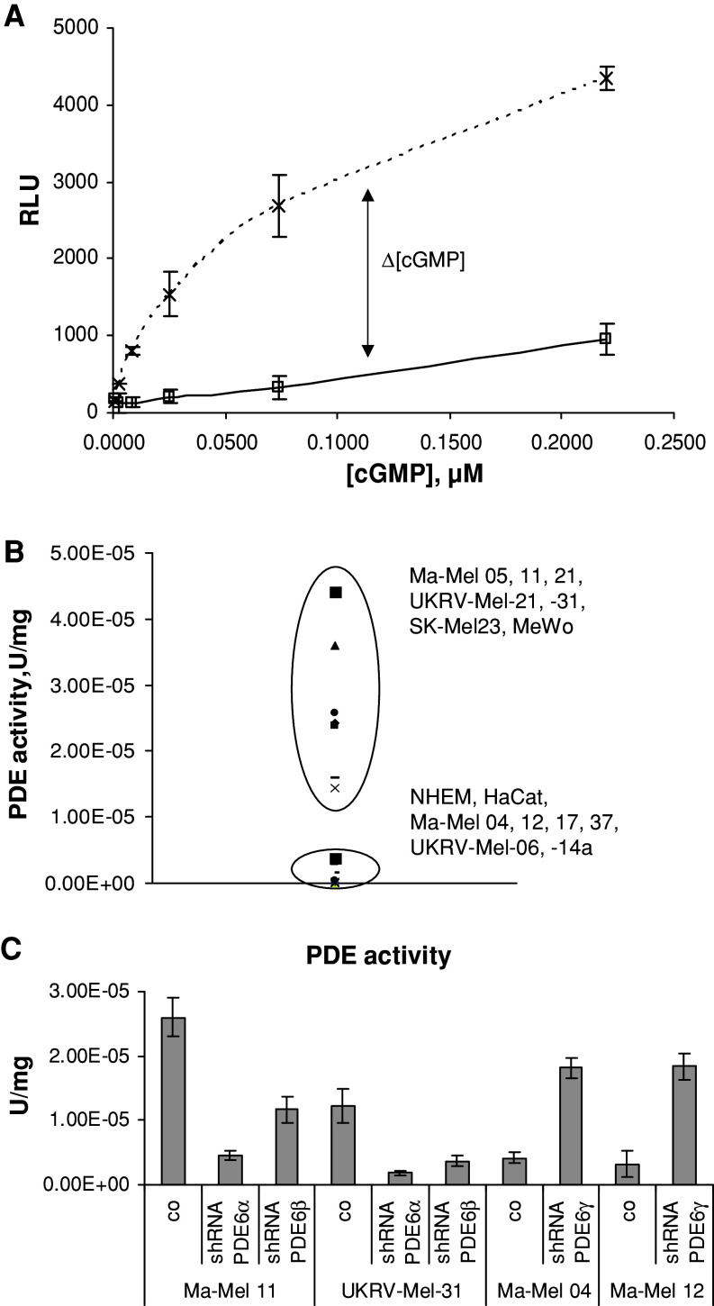
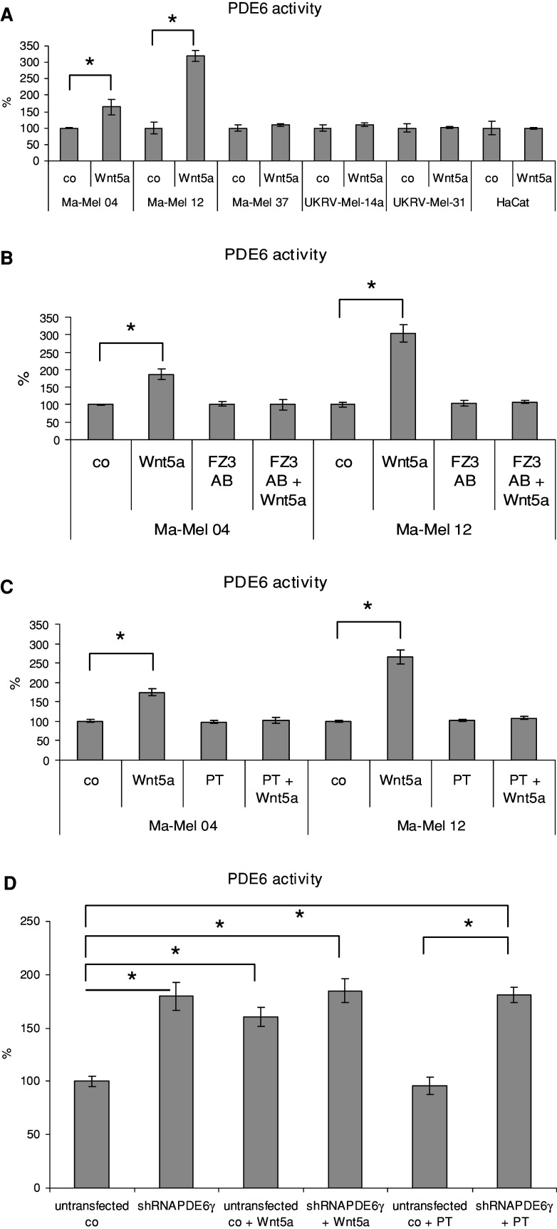
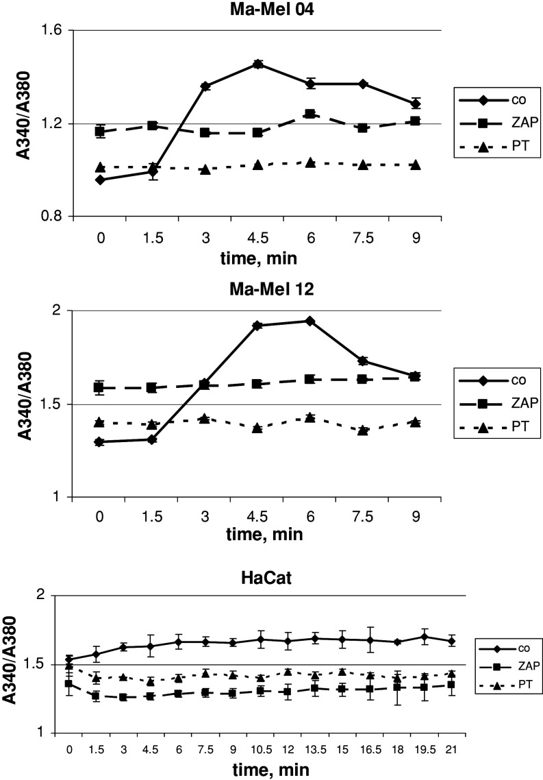
Malignant melanoma is one of the most aggressive human neoplasms which develop from the malignant transformation of normal epithelial melanocytes and share the lineage with retinal cells. cGMP-phosphodiesterase 6 (PDE6) is one of the cancer-retina antigens newly identified in melanoma cells. Normally, PDE6 hydrolyzes the photoreceptor second messenger cGMP allowing the visual signal transduction in photoreceptor cells. cGMP also play an important signaling role in stimulating melanogenesis in human melanocytes. Here, we present evidence that PDE6 is a key enzyme regulating the cGMP metabolism in melanoma cells. Decrease in intracellular cGMP leads to calcium accumulation in melanoma cells. In these cells, cGMP-phosphodiesterase 6 can be activated by another cancer-retina antigen, transducin, through Wnt5a-Frizzled-2 cascade, which leads to a lowering of cGMP and an increase in intracellular calcium mobilization. Thus, the aberrant expression of PDE6 may control cGMP metabolism and calcium homeostasis in melanoma cells.
Figures


Similar articles
-
Suppression of cyclic GMP-dependent protein kinase is essential to the Wnt/cGMP/Ca2+ pathway.J Biol Chem. 2006 Oct 13;281(41):30990-1001. doi: 10.1074/jbc.M603603200. Epub 2006 Aug 18. J Biol Chem. 2006. PMID: 16920709
-
Mitogen-activated protein kinase p38 regulates the Wnt/cyclic GMP/Ca2+ non-canonical pathway.J Biol Chem. 2007 Sep 28;282(39):28980-28990. doi: 10.1074/jbc.M702840200. Epub 2007 Aug 7. J Biol Chem. 2007. PMID: 17684012
-
WNT-frizzled signaling via cyclic GMP.Front Biosci. 2004 May 1;9:1043-7. doi: 10.2741/1310. Front Biosci. 2004. PMID: 14977527 Review.
-
Functional mapping of interacting regions of the photoreceptor phosphodiesterase (PDE6) γ-subunit with PDE6 catalytic dimer, transducin, and regulator of G-protein signaling9-1 (RGS9-1).J Biol Chem. 2012 Jul 27;287(31):26312-20. doi: 10.1074/jbc.M112.377333. Epub 2012 Jun 4. J Biol Chem. 2012. PMID: 22665478 Free PMC article.
-
Hear the Wnt Ror: how melanoma cells adjust to changes in Wnt.Pigment Cell Melanoma Res. 2009 Dec;22(6):724-39. doi: 10.1111/j.1755-148X.2009.00627.x. Epub 2009 Aug 25. Pigment Cell Melanoma Res. 2009. PMID: 19708915 Free PMC article. Review.
Cited by
-
Wnt signaling and the control of human stem cell fate.Stem Cell Rev Rep. 2014 Apr;10(2):207-29. doi: 10.1007/s12015-013-9486-8. Stem Cell Rev Rep. 2014. PMID: 24323281 Review.
-
WNT2-Mediated FZD2 Stabilization Regulates Esophageal Cancer Metastasis via STAT3 Signaling.Front Oncol. 2020 Jul 16;10:1168. doi: 10.3389/fonc.2020.01168. eCollection 2020. Front Oncol. 2020. PMID: 32766155 Free PMC article.
-
Signaling components of the 1α,25(OH)2D3-dependent Pdia3 receptor complex are required for Wnt5a calcium-dependent signaling.Biochim Biophys Acta. 2014 Nov;1843(11):2365-75. doi: 10.1016/j.bbamcr.2014.06.006. Epub 2014 Jun 16. Biochim Biophys Acta. 2014. PMID: 24946135 Free PMC article.
-
Phosphodiesterase Inhibitors as a Therapeutic Approach to Neuroprotection and Repair.Int J Mol Sci. 2017 Mar 24;18(4):696. doi: 10.3390/ijms18040696. Int J Mol Sci. 2017. PMID: 28338622 Free PMC article. Review.
-
Frizzled homolog proteins, microRNAs and Wnt signaling in cancer.Int J Cancer. 2013 Apr 15;132(8):1731-40. doi: 10.1002/ijc.27746. Epub 2012 Aug 30. Int J Cancer. 2013. PMID: 22833265 Free PMC article. Review.
References
Publication types
MeSH terms
Substances
LinkOut - more resources
Full Text Sources
Medical
Molecular Biology Databases
Miscellaneous
