

Positron emission tomography imaging of prostate cancer
- PMID: 19946787
- PMCID: PMC2883014
- DOI: 10.1007/s00726-009-0394-9
Positron emission tomography imaging of prostate cancer
Abstract
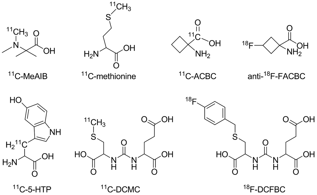
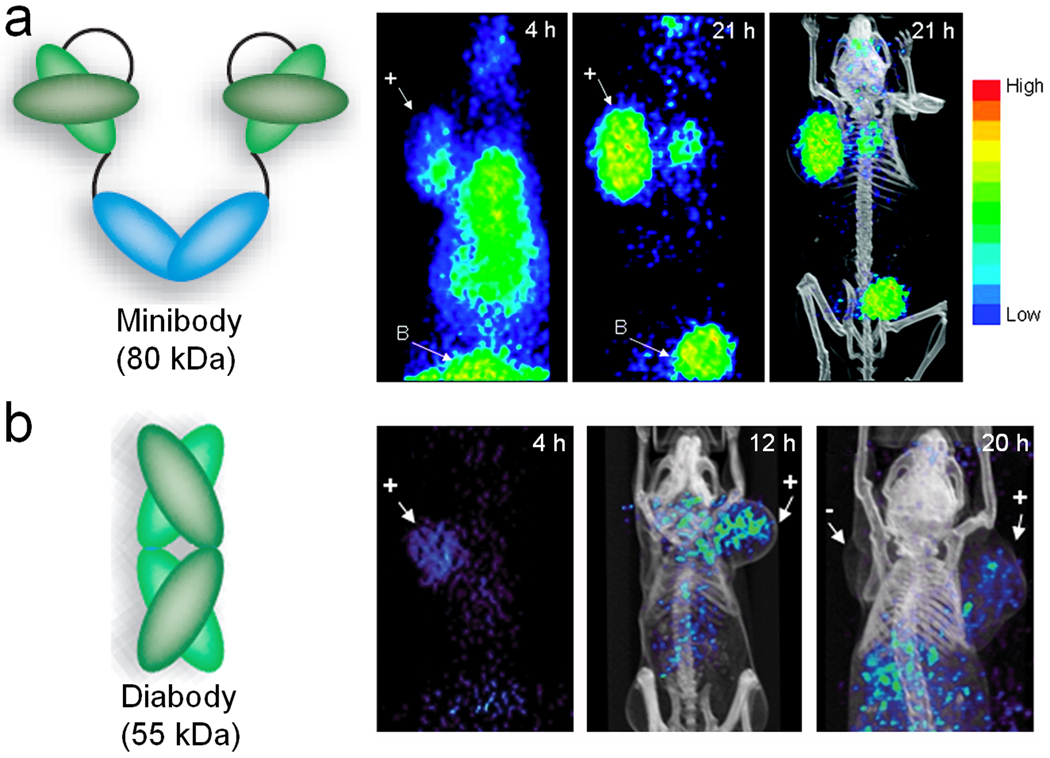
Prostate cancer (PCa) is the second leading cause of cancer death among men in the United States. Positron emission tomography (PET), a non-invasive, sensitive, and quantitative imaging technique, can facilitate personalized management of PCa patients. There are two critical needs for PET imaging of PCa, early detection of primary lesions and accurate imaging of PCa bone metastasis, the predominant cause of death in PCa. Because the most widely used PET tracer in the clinic, (18)F-fluoro-2-deoxy-2-D-glucose ((18)F-FDG), does not meet these needs, a wide variety of PET tracers have been developed for PCa imaging that span an enormous size range from small molecules to intact antibodies. In this review, we will first summarize small-molecule-based PET tracers for PCa imaging, which measure certain biological events, such as cell membrane metabolism, fatty acid synthesis, and receptor expression. Next, we will discuss radiolabeled amino acid derivatives (e.g. methionine, leucine, tryptophan, and cysteine analogs), which are primarily based on the increased amino acid transport of PCa cells. Peptide-based tracers for PET imaging of PCa, mostly based on the bombesin peptide and its derivatives which bind to the gastrin-releasing peptide receptor, will then be presented in detail. We will also cover radiolabeled antibodies and antibody fragments (e.g. diabodies and minibodies) for PET imaging of PCa, targeting integrin alpha(v)beta(3), EphA2, the epidermal growth factor receptor, or the prostate stem cell antigen. Lastly, we will identify future directions for the development of novel PET tracers for PCa imaging, which may eventually lead to personalized management of PCa patients.
Figures


Similar articles
-
More advantages in detecting bone and soft tissue metastases from prostate cancer using 18F-PSMA PET/CT.Hell J Nucl Med. 2019 Jan-Apr;22(1):6-9. doi: 10.1967/s002449910952. Epub 2019 Mar 7. Hell J Nucl Med. 2019. PMID: 30843003
-
Radionuclide based imaging of prostate cancer.Curr Top Med Chem. 2010;10(16):1600-16. doi: 10.2174/156802610793176774. Curr Top Med Chem. 2010. PMID: 20583988 Review.
-
PET Tracers Beyond FDG in Prostate Cancer.Semin Nucl Med. 2016 Nov;46(6):507-521. doi: 10.1053/j.semnuclmed.2016.07.005. Epub 2016 Sep 7. Semin Nucl Med. 2016. PMID: 27825431 Free PMC article. Review.
-
A comparative study of peptide-based imaging agents [68Ga]Ga-PSMA-11, [68Ga]Ga-AMBA, [68Ga]Ga-NODAGA-RGD and [68Ga]Ga-DOTA-NT-20.3 in preclinical prostate tumour models.Nucl Med Biol. 2020 May-Jun;84-85:88-95. doi: 10.1016/j.nucmedbio.2020.03.005. Epub 2020 Mar 30. Nucl Med Biol. 2020. PMID: 32251995
-
A Prospective Comparison of 18F-prostate-specific Membrane Antigen-1007 Positron Emission Tomography Computed Tomography, Whole-body 1.5 T Magnetic Resonance Imaging with Diffusion-weighted Imaging, and Single-photon Emission Computed Tomography/Computed Tomography with Traditional Imaging in Primary Distant Metastasis Staging of Prostate Cancer (PROSTAGE).Eur Urol Oncol. 2021 Aug;4(4):635-644. doi: 10.1016/j.euo.2020.06.012. Epub 2020 Jul 13. Eur Urol Oncol. 2021. PMID: 32675047 Clinical Trial.
Cited by
-
Prostate cancer metastases alter bone mineral and matrix composition independent of effects on bone architecture in mice--a quantitative study using microCT and Raman spectroscopy.Bone. 2013 Oct;56(2):454-60. doi: 10.1016/j.bone.2013.07.006. Epub 2013 Jul 15. Bone. 2013. PMID: 23867219 Free PMC article.
-
Image Guided Planning for Prostate Carcinomas With Incorporation of Anti-3-[18F]FACBC (Fluciclovine) Positron Emission Tomography: Workflow and Initial Findings From a Randomized Trial.Int J Radiat Oncol Biol Phys. 2016 Sep 1;96(1):206-13. doi: 10.1016/j.ijrobp.2016.04.023. Epub 2016 Apr 30. Int J Radiat Oncol Biol Phys. 2016. PMID: 27511856 Free PMC article. Clinical Trial.
-
Sepia ink oligopeptide induces apoptosis in prostate cancer cell lines via caspase-3 activation and elevation of Bax/Bcl-2 ratio.Mar Drugs. 2012 Oct;10(10):2153-2165. doi: 10.3390/md10102153. Epub 2012 Sep 27. Mar Drugs. 2012. PMID: 23170075 Free PMC article.
-
Detection of recurrent prostate carcinoma with anti-1-amino-3-18F-fluorocyclobutane-1-carboxylic acid PET/CT and 111In-capromab pendetide SPECT/CT.Radiology. 2011 Jun;259(3):852-61. doi: 10.1148/radiol.11102023. Epub 2011 Apr 14. Radiology. 2011. PMID: 21493787 Free PMC article.
-
Advancement in treatment and diagnosis of pancreatic cancer with radiopharmaceuticals.World J Gastrointest Oncol. 2016 Feb 15;8(2):165-72. doi: 10.4251/wjgo.v8.i2.165. World J Gastrointest Oncol. 2016. PMID: 26909131 Free PMC article. Review.
References
-
- Al-Nahhas A, Win Z, Szyszko T, Singh A, Khan S, Rubello D. What can gallium-68 PET add to receptor and molecular imaging? Eur J Nucl Med Mol Imaging. 2007;34:1897–1901. - PubMed
-
- Albrecht S, Buchegger F, Soloviev D, Zaidi H, Vees H, Khan HG, Keller A, Bischof Delaloye A, Ratib O, Miralbell R. 11C-acetate PET in the early evaluation of prostate cancer recurrence. Eur J Nucl Med Mol Imaging. 2007;34:185–196. - PubMed
-
- Ananias HJ, de Jong IJ, Dierckx RA, van de Wiele C, Helfrich W, Elsinga PH. Nuclear imaging of prostate cancer with gastrin-releasing-peptide-receptor targeted radiopharmaceuticals. Curr Pharm Des. 2008;14:3033–3047. - PubMed
Publication types
MeSH terms
Grants and funding
LinkOut - more resources
Full Text Sources
Other Literature Sources
Medical
Research Materials
Miscellaneous
