

Neuropsychiatric correlates of white matter hyperintensities in Alzheimer's disease
- PMID: 19946864
- PMCID: PMC3975914
- DOI: 10.1002/gps.2418
Neuropsychiatric correlates of white matter hyperintensities in Alzheimer's disease
Abstract
Objective: To investigate the association of behavioral and psychological symptoms of dementia (BPSD) in Alzheimer's disease (AD) and magnetic resonance imaging (MRI) measures of brain atrophy and white matter hyperintensities (WMH).
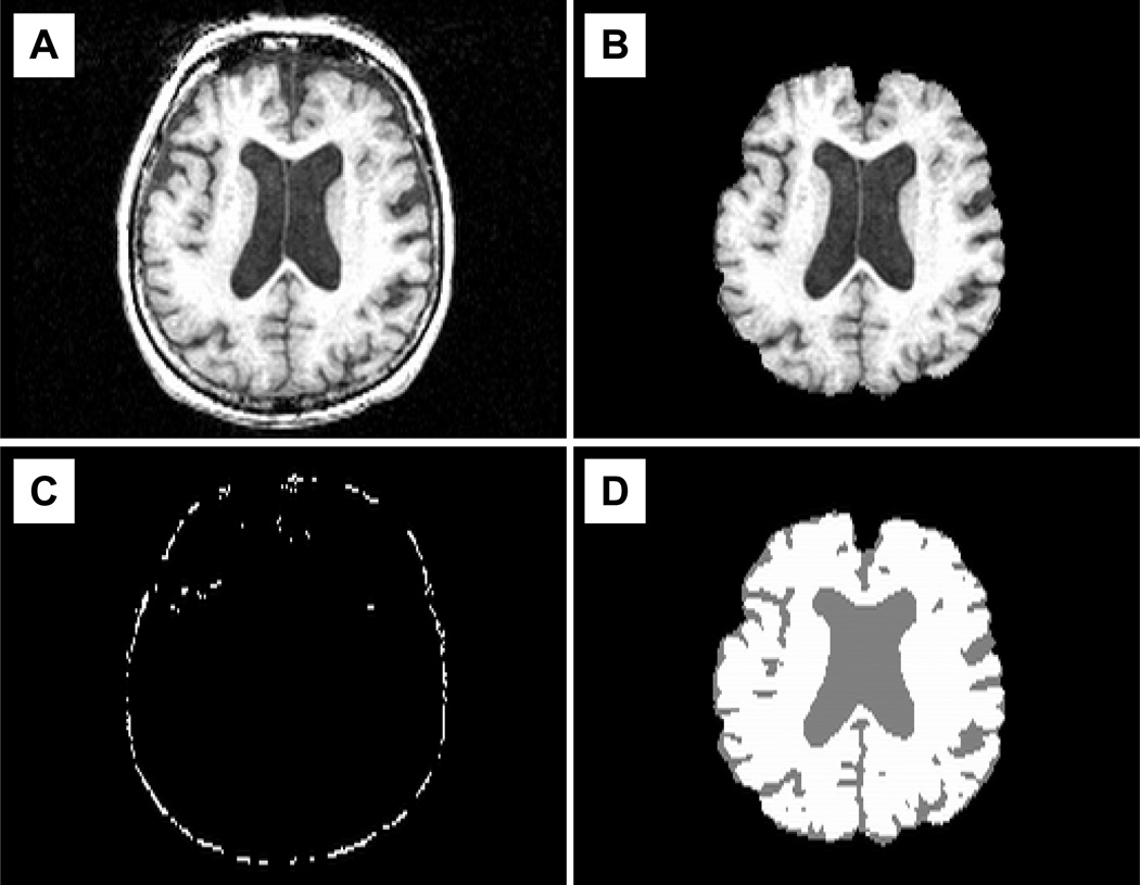
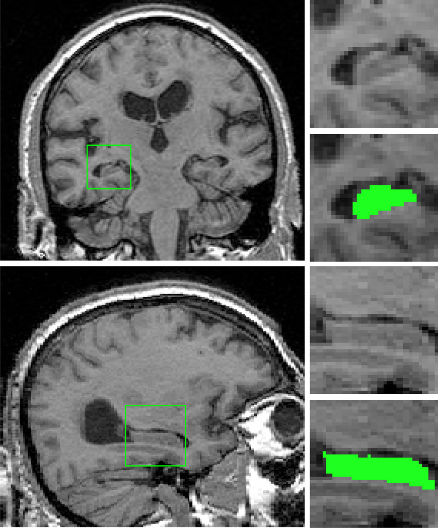
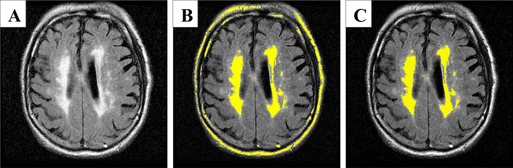
Methods: Thirty-seven patients with probable AD received the Neuropsychiatric Inventory (NPI), the Mini Mental Status Exam (MMSE), and an MRI scan as part of their initial evaluation at the Outpatient Memory Diagnostic Clinic at McLean Hospital. MRI-based volumetric measurements of whole brain atrophy, hippocampal volumes, and WMH were obtained. Analysis of covariance models, using age as a covariate and the presence of specific BPSD as independent variables, were used to test for differences in whole brain volumes, hippocampal volumes and WMH volumes.
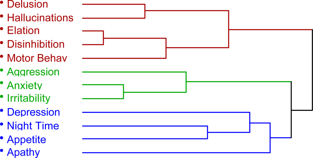
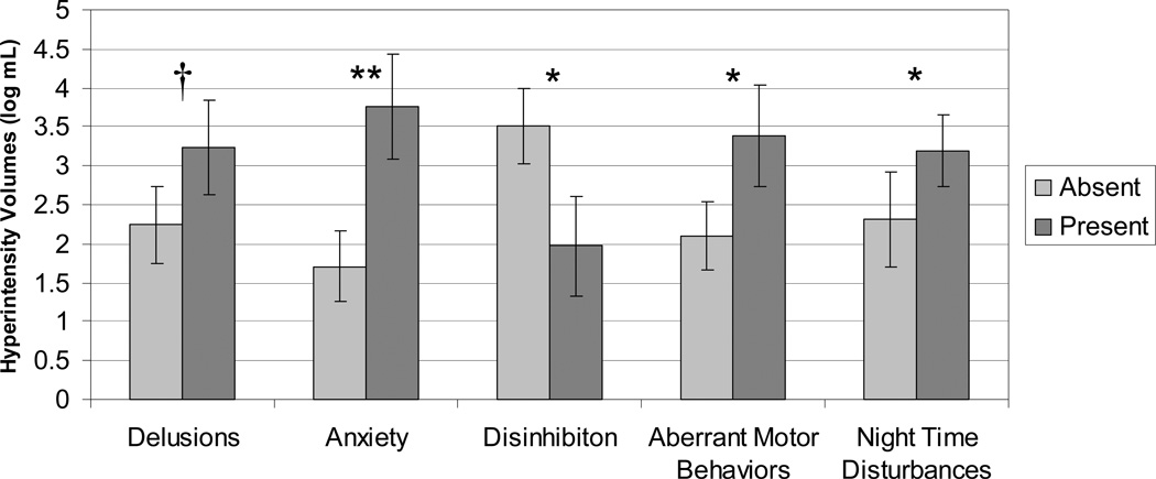
Results: Increased WMH were associated with symptoms of anxiety, aberrant motor behavior, and night time disturbance, while symptoms of disinhibition were linked to lower WMH volume. No associations were found for whole brain or hippocampal volumes and BPSD.
Conclusions: These findings suggest that white matter changes are associated with the presence of BPSD in AD.
Figures
Similar articles
-
Medial temporal lobe atrophy is independently associated with behavioural and psychological symptoms in Alzheimer's disease.Psychogeriatrics. 2019 Jan;19(1):46-54. doi: 10.1111/psyg.12363. Epub 2018 Aug 6. Psychogeriatrics. 2019. PMID: 30084177
-
Evaluation of AccuBrain-based MRI quantitative analysis in diagnosing Alzheimer's disease and assessing behavioral and psychological symptoms of dementia.Aging Clin Exp Res. 2025 Apr 17;37(1):126. doi: 10.1007/s40520-025-03023-6. Aging Clin Exp Res. 2025. PMID: 40244500 Free PMC article.
-
Reconsidering harbingers of dementia: progression of parietal lobe white matter hyperintensities predicts Alzheimer's disease incidence.Neurobiol Aging. 2015 Jan;36(1):27-32. doi: 10.1016/j.neurobiolaging.2014.07.019. Epub 2014 Jul 21. Neurobiol Aging. 2015. PMID: 25155654 Free PMC article.
-
White matter hyperintensities are significantly associated with cortical atrophy in Alzheimer's disease.J Neurol Neurosurg Psychiatry. 2004 Jun;75(6):822-7. doi: 10.1136/jnnp.2003.019273. J Neurol Neurosurg Psychiatry. 2004. PMID: 15145992 Free PMC article.
-
[White Matter Lesion and Alzheimer's Disease: The Association between Small Vessel Disease and Neuropsychiatric Symptoms in Alzheimer's Disease].Brain Nerve. 2015 Apr;67(4):427-32. doi: 10.11477/mf.1416200158. Brain Nerve. 2015. PMID: 25846591 Review. Japanese.
Cited by
-
Depressive Symptoms Have Distinct Relationships With Neuroimaging Biomarkers Across the Alzheimer's Clinical Continuum.Front Aging Neurosci. 2022 Jun 20;14:899158. doi: 10.3389/fnagi.2022.899158. eCollection 2022. Front Aging Neurosci. 2022. PMID: 35795235 Free PMC article.
-
Neurobiology of neuropsychiatric symptoms in Alzheimer's disease: A critical review with a focus on neuroimaging.Dement Neuropsychol. 2013 Jul-Sep;7(3):236-243. doi: 10.1590/S1980-57642013DN70300002. Dement Neuropsychol. 2013. PMID: 29213845 Free PMC article. Review.
-
Frontal White Matter Hyperintensity Is Associated with Verbal Aggressiveness in Elderly Women with Alzheimer Disease and Amnestic Mild Cognitive Impairment.Dement Geriatr Cogn Dis Extra. 2018 Apr 11;8(1):138-150. doi: 10.1159/000486826. eCollection 2018 Jan-Apr. Dement Geriatr Cogn Dis Extra. 2018. PMID: 29805380 Free PMC article.
-
The Relationship Between Anxiety and Alzheimer's Disease.J Alzheimers Dis Rep. 2021 Mar 8;5(1):171-177. doi: 10.3233/ADR-210294. J Alzheimers Dis Rep. 2021. PMID: 33981954 Free PMC article. Review.
-
Association of Cerebrovascular Imaging Biomarkers, Depression, and Anxiety, with Mild Cognitive Impairment.J Alzheimers Dis Rep. 2023 Nov 3;7(1):1237-1246. doi: 10.3233/ADR-230073. eCollection 2023. J Alzheimers Dis Rep. 2023. PMID: 38025797 Free PMC article.
References
-
- Aharon-Peretz J, Masiah A, Pillar T, Epstein R, Tzischinsky O, Lavie P. Sleep-wake cycles in multi-infarct dementia and dementia of the Alzheimer type. Neurology. 1991;41(10):1616–1619. lost in the final stages of Alzheimer's disease. - PubMed
-
- Assal F, Cummings JL. Neuropsychiatric symptoms in the dementias. Curr Opin Neurol. 2002;15(4):445–450. - PubMed
-
- Association AP. Diagnostic and Statistical Manual of Mental Disorders. (4th ed.) Washington D.C: American Psychiatric Association; 1994.
-
- Ballard C, Neill D, O'Brien J, McKeith IG, Ince P, Perry R. Anxiety, depression and psychosis in vascular dementia: prevalence and associations. J Affect Disord. 2000;59(2):97–106. - PubMed
Publication types
MeSH terms
Grants and funding
LinkOut - more resources
Full Text Sources
Medical
