

Isolation and characterization of a strain of Lichtheimia corymbifera (ex Absidia corymbifera) from a case of bovine abortion
- PMID: 19948023
- PMCID: PMC2789724
- DOI: 10.1186/1477-7827-7-138
Isolation and characterization of a strain of Lichtheimia corymbifera (ex Absidia corymbifera) from a case of bovine abortion
Abstract
Background: Lichtheimia corymbifera (previously Absidia corymbifera) is a filamentous zygomycetes belonging to the order Mucorales and to the family Lichtheimiaceae. Members of genus Lichtheimia spp. are cosmopolitan and ubiquitous in nature. Lichtheimia corymbifera is a recognized agent of diseases in man and animals. In cattle it causes abortion and mastitis. Three cases of bovine abortion occurred in a herd located in the Po Valley. Serological examinations were performed on fetal and mother's blood. One of the aborted fetus was referred to our laboratory. The paper describes the isolation and characterization of Lichtheimia corymbifera from a bovine aborted fetus.
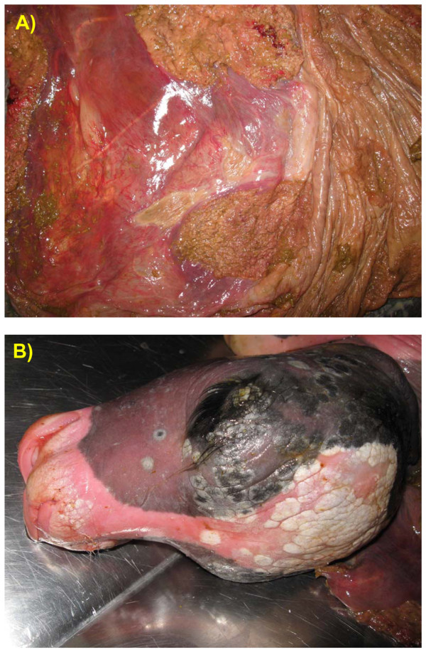
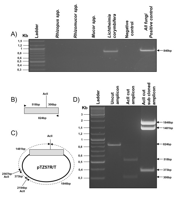
Methods: Serological examinations were performed on fetal and mother's blood. Lesions on fetal tissues and placenta leaded the diagnostic suspect towards a mycotic aetiology. Tissues were then put in culture, and at the same time an histological examination was performed, together with bacteriological and virological tests. The isolate from placenta and fetal tissues was identified and characterized by PCR and RFLP, using the ITS region as a target sequence and AclI restriction site within the amplicon to distinguish Lichtheimia corymbifera among the other fungi.
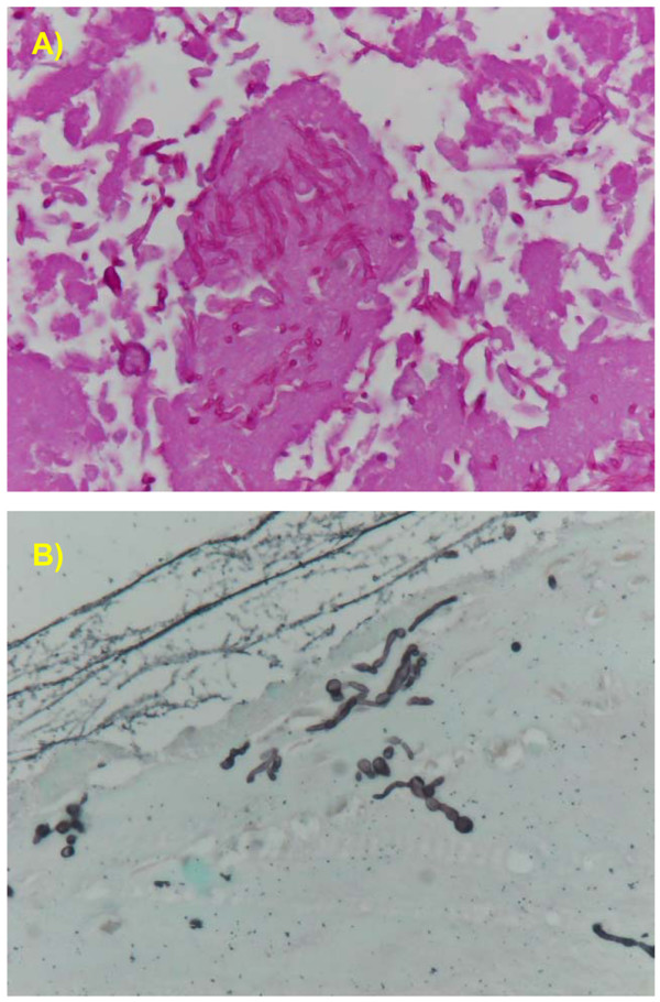
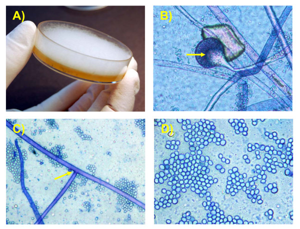
Results: Serological, bacteriological and virological tests gave aspecific results. Histological examination evidenced numerous PAS positive hyphae within the necrotic cotiledons and numerous fungal nonseptate hyphae to the GMS stain. Colonies with typical morphological features of fungi grew up on Sabouraud agar from fetal skin and placenta. On the developed colonies the microscopic examination has shown a large number of nonseptate hyphae and sporangia consistent with Mucorales. PCR and RFLP allowed the identification of the isolate as Lichtheimia corymbifera.
Conclusion: The present report describes the isolation and the molecular characterisation of a fungal isolate from bovine aborted fetus and placenta. The diagnostic protocol allowed to identify and characterise the strain. This is the first isolation in Italy of Lichtheimia corymbifera in a bovine aborted fetus.
Figures
References
-
- Committee on Bovine Reproductive Nomenclature. Recommendations for Standardizing Bovine Reproduction Terms. Cornell Vet. 1972;62:216–237. - PubMed
-
- Anderson ML. Infectious causes of bovine abortion during mid-to late-gestation. Theriogenology. 2007;68:474–486. - PubMed
-
- Knudtson WU, Kirkbride CA. Fungi associated with bovine abortion in the northern plains states (USA) J Vet Diagn Invest. 1992;4:181–185. - PubMed
-
- Ali R, Khan IH. Mycotic abortion in cattle. Pak Vet J. 2006;26:44–46.
Publication types
MeSH terms
LinkOut - more resources
Full Text Sources
Medical
Miscellaneous
