. 2009 Nov 30:9:59.
doi: 10.1186/1471-213X-9-59.

Zebrafish con/disp1 reveals multiple spatiotemporal requirements for Hedgehog-signaling in craniofacial development

Affiliations

Zebrafish con/disp1 reveals multiple spatiotemporal requirements for Hedgehog-signaling in craniofacial development

Tyler Schwend et al. BMC Dev Biol. .

Abstract

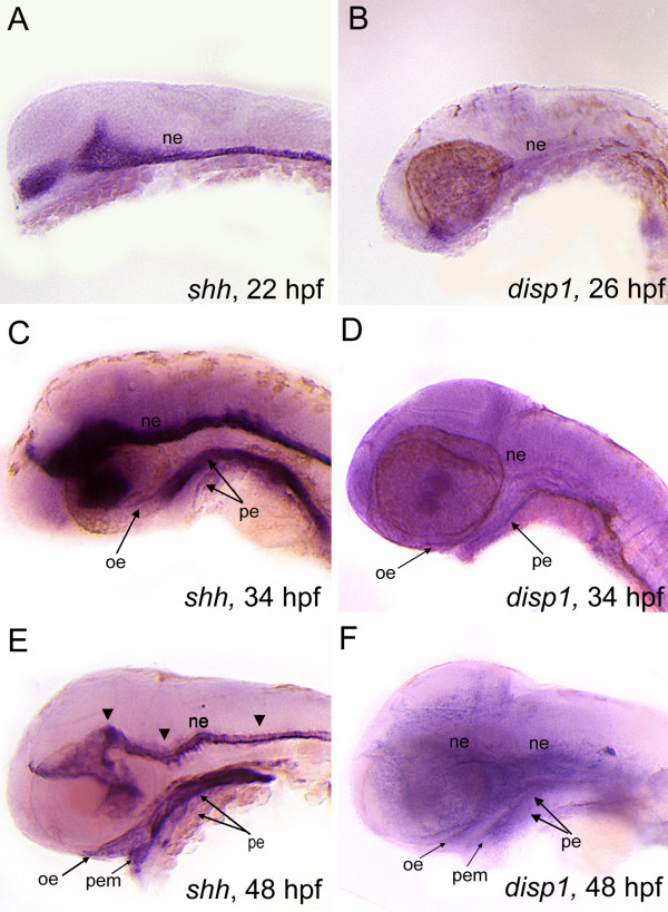
Background: The vertebrate head skeleton is derived largely from cranial neural crest cells (CNCC). Genetic studies in zebrafish and mice have established that the Hedgehog (Hh)-signaling pathway plays a critical role in craniofacial development, partly due to the pathway's role in CNCC development. Disruption of the Hh-signaling pathway in humans can lead to the spectral disorder of Holoprosencephaly (HPE), which is often characterized by a variety of craniofacial defects including midline facial clefting and cyclopia 12. Previous work has uncovered a role for Hh-signaling in zebrafish dorsal neurocranium patterning and chondrogenesis, however Hh-signaling mutants have not been described with respect to the ventral pharyngeal arch (PA) skeleton. Lipid-modified Hh-ligands require the transmembrane-spanning receptor Dispatched 1 (Disp1) for proper secretion from Hh-synthesizing cells to the extracellular field where they act on target cells. Here we study chameleon mutants, lacking a functional disp1(con/disp1).

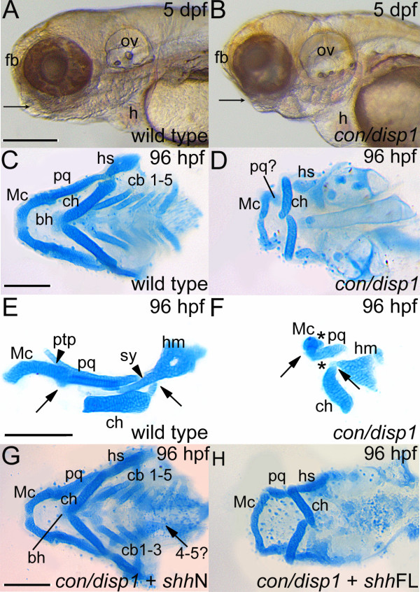
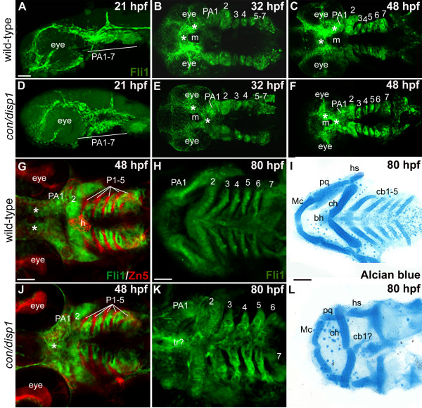
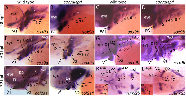
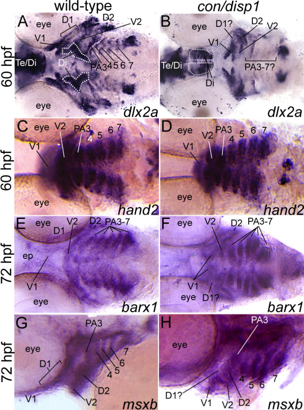
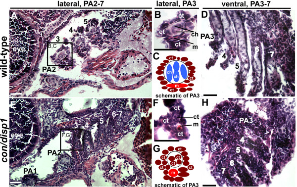
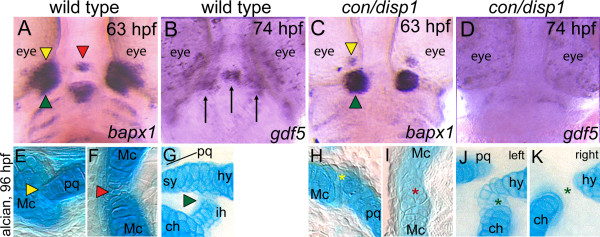
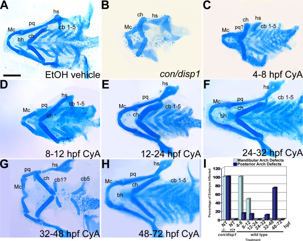
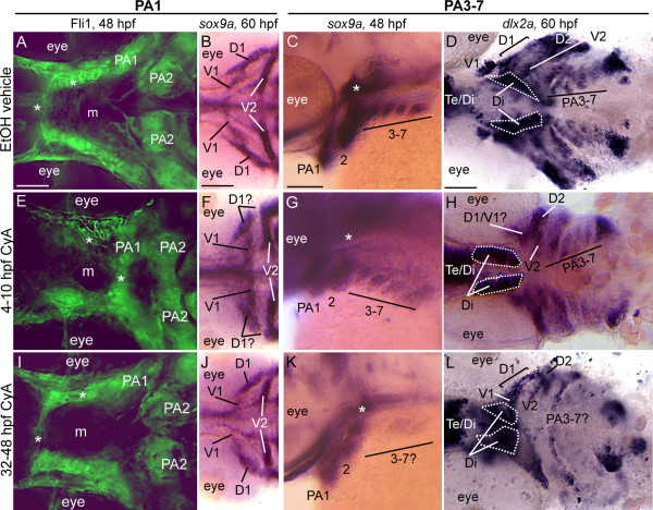
Results: con/disp1 mutants display reduced and dysmorphic mandibular and hyoid arch cartilages and lack all ceratobranchial cartilage elements. CNCC specification and migration into the PA primorida occurs normally in con/disp1 mutants, however disp1 is necessary for post-migratory CNCC patterning and differentiation. We show that disp1 is required for post-migratory CNCC to become properly patterned within the first arch, while the gene is dispensable for CNCC condensation and patterning in more posterior arches. Upon residing in well-formed pharyngeal epithelium, neural crest condensations in the posterior PA fail to maintain expression of two transcription factors essential for chondrogenesis, sox9a and dlx2a, yet continue to robustly express other neural crest markers. Histology reveals that posterior arch residing-CNCC differentiate into fibrous-connective tissue, rather than becoming chondrocytes. Treatments with Cyclopamine, to inhibit Hh-signaling at different developmental stages, show that Hh-signaling is required during gastrulation for normal patterning of CNCC in the first PA, and then during the late pharyngula stage, to promote CNCC chondrogenesis within the posterior arches. Further, loss of disp1 disrupted normal expression of bapx1 and gdf5, markers of jaw joint patterning, thus resulting in jaw joint defects in con/disp1 mutant animals.

Conclusion: This study reveals novel requirements for Hh-signaling in the zebrafish PA skeleton and highlights the functional diversity and differential sensitivity of craniofacial tissues to Hh-signaling throughout the face, a finding that may help to explain the spectrum of human facial phenotypes characteristic of HPE.


Figures

Figure 1
Figure 1
con/disp1 mutants display defective cartilage development in the PA which is partly rescued by RNA encoding non lipid-modified Shh. (A,B) Lateral views of live larvae show a reduction in head tissue and a deficit in jaw outgrowth (arrowhead) in con/disp1 mutant larvae at 5 dpf. (C) Ventral view of 96 hpf Alcian blue-stained wild type cartilages. (D) con/disp1 mutant cartilages reveal reduced, hypoplastic mandibular and hyoid arch cartilage elements and a complete absence of the cb 1-5 elements and midline-forming bh cartilage. (E,F) Lateral view of jaw cartilages reveals malformed joints (arrows denote joint site), fewer chondrocytes contributing to the pq cartilage element and a complete loss of the ptp and sy cartilage element in con/disp1 mutant (arrowhead denotes presence of cartilage, asterisk denotes cartilage absence). (G) Injection of 150 pg shhN mRNA into genotype-confirmed con/disp1 mutant mostly restores cartilage elements to wild type state (arrow indicates unilateral rescue in cb 4-5 elements), while (H) con/disp1 mutant injected with 150 pg shhFL mRNA are phenotypically similar to noninjected con/disp1 larvae. Scale bar: 50 μM.
Figure 2
Figure 2
CNCC patterning in the con/disp1 mutant is visualized by the fli1gfp transgene. Lateral views (A,D) or ventral views (B,C,E-L) of confocal stack projections of fli1GFP+ CNCC in wild type and con/disp1 mutants (A-H, J,K) or Alcian blue-stained larvae to visualize cartilages (I,L). (G,J) Zn5 (red) staining to visualize endodermal pouches (P 1-5) in fli1GFP embryos. (A,D) At 21 hpf, wild type (A) and con/disp1 mutants (D) reveal similar patterns of postmigratory-CNCC. (B,C) 32 hpf ventral views of wild type embryos reveal CNC condensations in PA (B), and segmentation of CNCC by 48 hpf (C). (E,F) By 32 hpf, con/disp1 anterior-most CNCC (asterisks) become mispatterned, while CNCC within more PA 2-7 are correctly patterned (E) and segmented by 48 hpf (F). (G,J) CNCC in wild type (G) and con/disp1 mutants (J) are interdigitated properly in well-formed endodermal pouches 1-5 (P 1-5) (Zn5, red). (H,K) con/disp1 mutants show developed, segmented CNCC in posterior arches (K). (I,L) CNCC within con/disp1 mutant posterior arches fail to become cartilage. Scale bar: 50 μM.
Figure 3
Figure 3
Chondrogenic differentiation in con/disp1 CNCC. Lateral views of 48 hpf (A-D), 60 hpf (E-H), 72 hpf (I,J) or ventral views of 72 hpf (K,L) wild type (A,C,E,G,I,K) or con/disp1 (B,D,F,H,J,L) embryos labeled with RNA probe for sox9a (A,B,E,F), sox9b (C,D,G,H), col2a1 (I,J) or runx2b (K,L). (A,C) sox9a marks CNCC in PA1-7 at 48 hpf in wild type embryos (A), however expression is lost in the posterior arch-residing CNCC in con/disp1 mutants (E). (C,D) sox9b continues to be expressed in 48 hpf PA1-7 condensations in wild type (B) and con/disp1 mutants alike (D). (E-H) At 60 hpf, sox9a and sox9b becomes downregulated in the dorsal subset of con/disp1 CNCC in the anterior arches (D1 and D2). sox9a remains absent in posterior arches in con/disp1 mutants, while sox9b remains robustly expressed in posterior arches. (I,J) At 72 hpf, col2a1 is reduced in con/disp1 D1 and D2 condensations and absent in PA3-7. (K,L) At 72 hpf, runx2b is absent in con/disp1 D1 and dorsal trabeculae precursors, but is present in PA condensation at wild type levels. (K',L') insets are of lateral views of same embryo shown in (K) and (L) respectively. Lateral views show presence of runx2b expression in opercle condensation in con/disp1 and wild type embryos alike. Asterisks indicate expression in mesoderm-derived polar cartilages. Images are at same magnification.
Figure 4
Figure 4
con/disp1 mutants show variable postmigratory-CNCC gene expression defects. Ventral, 60 hpf (A-D), ventral, 72 hpf (E,F) or lateral, 72 hpf (G,H) views of wild type (A,C,E,G) or con/disp1 (B,D,F,H) embryos labeled with RNA probe for dlx2a (A,B), hand2 (C,D), barx1 (E,F), msxb (G,H). (A,B) dlx2a is downregulated in dorsal domain of PA1 (D1) and posterior arches in con/disp1 mutants. (C,D) hand2 is expressed in CNCC within each con/disp1 PA. (E,F) At 72 hpf, barx1 expression marks the anterior and posterior arches in wild type embryos (E), but is downregulated in the con/disp1 D1 domain of PA1 (F). (G,H) msxb is downregulated in the D1 domain, but is expressed at wild type levels in posterior arch-residing CNCC. Scale bar: 50 μM.
Figure 5
Figure 5
con/disp1 posterior arch-residing CNCC become fibrous-connective tissue. Lateral, (A,B,E,F) or ventral (D,H) H&E stained 10 μM sections of 5 dpf wild type (A,B,D) or con/disp1 mutant (E,F,H) larvae. (A,C) Chondrocytes are surrounded by connective tissue within each wild type PA (A), with higher magnification of PA3 revealing individual cell types (B). Box in (A) surrounding PA3 is viewed at higher magnification in (B). (E,F) con/disp1 mutants lack chondroctyes in PA3-7 (E), higher magnification of PA3 displays ectopic fibrous-connective tissue in PA3 (F). Box in (E) is viewed at higher magnification in (F). (C,G) Schematic of cell types visualized in wild type (C) and con/disp1 (G) PA3. (D,H) Horizontal sections show chondrocytes stacked within wild type posterior arches (D), while fibrous-connective tissue populate the con/disp1 posterior arches (H). Scale bar: 50 μM.
Figure 6
Figure 6
Joint defects are apparent in con/disp1 mutants. Ventral views (A-D) of wild-type (A,B) or con/disp1 mutants (C,D) labeled with RNA probe for bapx1 at 63 hpf (A,C) or gdf5 at 74 hpf (B,D). Alcian blue-stained cartilage at 96 hpf in wild-type (E-G) or con/disp1 mutant (H-K) embryos. (A,C) bapx1 prefigures bilateral and midline joints in wild-type embryos, but expression is reduced bilaterally and absent in the midline of con/disp1 mutants. Different color arrowheads used to denote individual cranial joints in A,C and E-G. Asterisks of the same color designate a reduced joint in con/disp1 mutants in H-K. (B,D) gdf5 is strongly expressed bilaterally in the first arch joint region (arrows) and in a group of cells in the midline that prefigures the basihyal cartilage element (arrow) (B). Expression of gdf5 is significantly reduced in both bilateral and midline domains in con/disp1 mutants (D). (E-G) In wild-type embryos, bilateral joints form between the Mc and pq (E, yellow arrowhead) and hs and ch (G, green arrowhead), as well as at the midline between the bilaterally formed Mc (F, red arrowhead). (H-K) In con/disp1 mutants, a poorly formed joint between the Mc, which fails to extend into a RAP element, and a severely reduced pq is visualized (H, yellow asterisk). In the second arch, we see either ectopic cartilage cells at the joint site between the hs and ch elements (J, green asterisk) or a failure of the hs and ch to join (K, green asterisk). Both phenotypes are occasionally present in the same larvae (left designates left side, right designates right side of single larvae). Further, the bilateral mc elements fuse at the midline to disrupt the joint (I, red asterisk).
Figure 7
Figure 7
shh and disp1 are coexpressedin the developing head. Lateral views of wild type embryos labeled with RNA probe for shh (A,C,E) or disp1 (B,D,F). (A,B) At 22 hpf (shh) - 25 hpf (disp1)are both expressed in ventral neuroectoderm (ne). (C,D) At 34 hpf, shh and disp1 expression becomes detectable in oral ectoderm (oe) and pharyngeal endoderm (pe), in addition to the neuroectoderm (ne). Arrowheads denote expression within the neuroectoderm for both shh and disp1. (E-F) By 48 hpf, shh and disp1 expression becomes more prominent in the oe and pe, and is further expanded to the pharyngeal ectodermal margin (pem). shh and disp1 expression persists in the ne at 48 hpf.
Figure 8
Figure 8
Early and late Cyclopamine treatments disrupt ventral PA development. (A-H) 96 hpf Alcian blue-stained cartilage. Wild type treated with ethanol (EtOH) vehicle (4-48 hpf) (A), untreated con/disp1 mutant (B) and wild type treated with 100 μM Cya at stages between 4-72 hpf (C-H). (B,C) 4-8 hpf treatment causes reduced jaw cartilage and outgrowth defects (C), similar to con/disp1 mutant (B), while hyoid and posterior arch cartilages are unaffected (C). (D-F) 8-32 hpf treatments had little effect on PA cartilage development, with occasional jaw cartilage reductions in 8-12 hpf treatment (D). (G) 32-48 hpf treatment eliminated most cb cartilages, without reducing anterior arch cartilage. (H) Treatments after 48 hpf have no effect on PA cartilage development. (I) Mandibular arch patterning defects and posterior arch chondrogenesis defects are summarized for each treatment (n = 20 embryos per treatment). Scale bar: 50 μM.
Figure 9
Figure 9
Early and late Cyclopamine treatments lead to CNCC defects. Ventral (A,B,D,E,F,H,I,J,L) or lateral (C,G,K) views at 48 hpf (A,C,E,G,I,K) or 60 hpf (B,D,F,H,J,L) of vehicle-treated wild type (A-D) or Cya-treated wild type (E-L) embryos that are Fli1gfp positive (A,E,I) or labeled with RNA probe for sox9a (B,C,F,G,J,K) or dlx2a (D,H,L). (A,E,I) By 48 hpf, vehicle-treated wild type embryos (A) display proper patterning of anteriormost CNCC (asterisks), as do 32-48 hpf Cya-treated embryos (I); however, 4-10 hpf Cya-treated embryos (E) show mispatterning of anteriormost CNCC. (B,F,J) Vehicle treated embryos display normal sox9a expression in the dorsal (D1) and ventral (V1) CNCC condensations in the first arch (B), as do 32-48 hpf Cya-treated embryos (J). In 4-10 hpf Cya-treated embryos, ventral (V1) CNCC condensations maintain sox9a expression, while sox9a is greatly reduced in the dorsal (D1) region of the first arch. (C,D,G,H,K,L) Treating wild type embryos with vehicle or Cya from 4-10 hpf does not influence sox9a expression at 48 hpf(C,G) or dlx2a at 60 hpf (D,H) within posterior arch residing CNCC, while treating wild type embryos with Cya at 32-48 hpf leads to reduction in sox9a gene expression at 48 hpf (K) and dlx2a at 60 hpf (L) within CNCC in the posterior arches. Asterisks in (C,G,K) indicate sox9a expression in mesoderm-derived polar cartilages. Scale bar: 50 μM.

Similar articles

Cited by

References

    1. Roessler E, Belloni E, Gaudenz K, Jay P, Berta P, Scherer SW, Tsui LC, Muenke M. Mutations in the human Sonic Hedgehog gene cause holoprosencephaly. Nat Genet. 1996;14:357–60. doi: 10.1038/ng1196-357. - DOI - PubMed
    1. Roessler E, Ma Y, Ouspenskaia MV, Lacbawan F, Bendavid C, Dubourg C, Beachy PA, Muenke M. Truncating loss-of-function mutations of DISP1 contribute to holoprosencephaly-like microform features in humans. Hum Genet. 2009;125:393–400. doi: 10.1007/s00439-009-0628-7. - DOI - PMC - PubMed
    1. Le Douarin NM. Cell migrations in embryos. Cell. 1984;38:353–60. doi: 10.1016/0092-8674(84)90490-2. - DOI - PubMed
    1. Couly G, Creuzet S, Bennaceur S, Vincent C, Le Douarin NM. Interactions between Hox-negative cephalic neural crest cells and the foregut endoderm in patterning the facial skeleton in the vertebrate head. Development. 2002;129:1061–73. - PubMed
    1. Noden DM. The embryonic origins of avian cephalic and cervical muscles and associated connective tissues. Am J Anat. 1983;168:257–76. doi: 10.1002/aja.1001680302. - DOI - PubMed

Publication types

Substances

LinkOut - more resources