. 2009 Dec 1:9:60.
doi: 10.1186/1471-213X-9-60.

Role of Scrib and Dlg in anterior-posterior patterning of the follicular epithelium during Drosophila oogenesis

Affiliations

Role of Scrib and Dlg in anterior-posterior patterning of the follicular epithelium during Drosophila oogenesis

Qi Li et al. BMC Dev Biol. .

Abstract

Background: Proper patterning of the follicle cell epithelium over the egg chamber is essential for the Drosophila egg development. Differentiation of the epithelium into several distinct cell types along the anterior-posterior axis requires coordinated activities of multiple signaling pathways. Previously, we reported that lethal(2)giant larvae (lgl), a Drosophila tumor suppressor gene, is required in the follicle cells for the posterior follicle cell (PFC) fate induction at mid-oogenesis. Here we explore the role of another two tumor suppressor genes, scribble (scrib) and discs large (dlg), in the epithelial patterning.

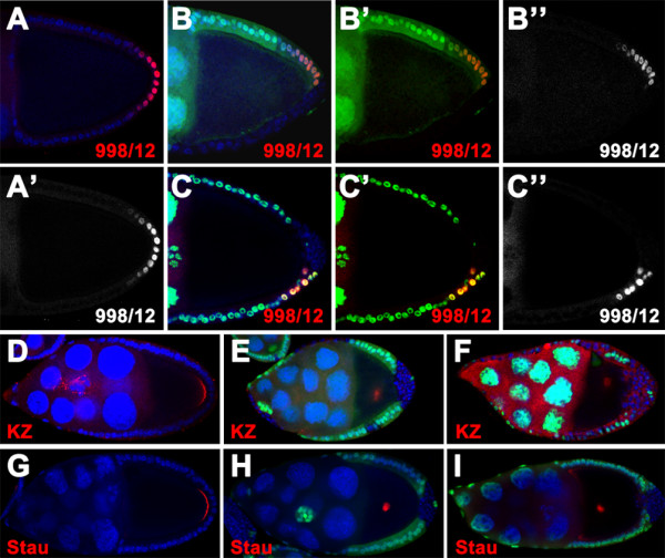
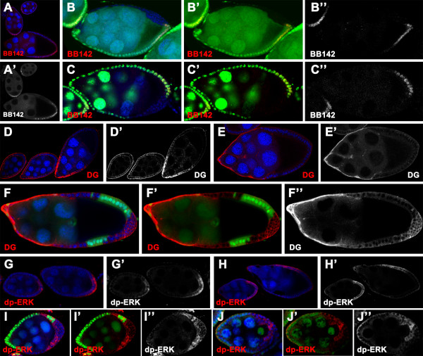
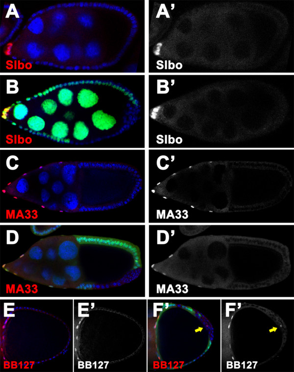
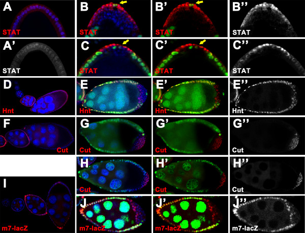
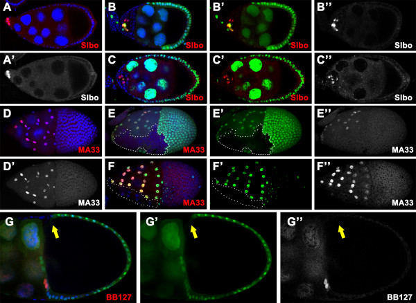
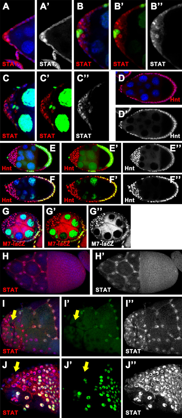
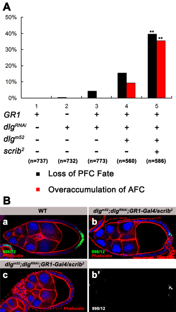
Results: We found that removal of scrib or dlg function from the follicle cells at posterior terminal of the egg chamber causes a complete loss of the PFC fate. Aberrant specification and differentiation of the PFCs in the mosaic clones can be ascribed to defects in coordinated activation of the EGFR, JAK and Notch signaling pathways in the multilayered cells. Meanwhile, the clonal analysis revealed that loss-of-function mutations in scrib/dlg at the anterior domains result in a partially penetrant phenotype of defective induction of the stretched and centripetal cell fate, whereas specification of the border cell fate can still occur in the most anterior region of the mutant clones. Further, we showed that scrib genetically interacts with dlg in regulating posterior patterning of the epithelium.

Conclusion: In this study we provide evidence that scrib and dlg function differentially in anterior and posterior patterning of the follicular epithelium at oogenesis. Further genetic analysis indicates that scrib and dlg act in a common pathway to regulate PFC fate induction. This study may open another window for elucidating role of scrib/dlg in controlling epithelial polarity and cell proliferation during development.


Figures

Figure 1
Figure 1
Loss of scrib or dlg function causes defects in specification and differentiation of the PFCs. Wild-type (A, D and G) and mutant egg chambers containing scrib2 (B, E and H) or dlgm52 (C, F and I) clones at the posterior marked by the absence of nuclear GFP (green in B, C, E, F and H) or β-gal (green in I), stained for nuclei (DAPI, blue) and β-gal(red in A-F) or Stau (red in G-I). (A-C) Expression of the PFC marker 998/12 can be observed in stage 10 wild type egg chambers (A, A'), whereas loss of 998/12 expression is evident in the scrib2 (B-B") and dlgm52 (C-C") clone cells. Note that 998/12 is still present in the remaining wild-type posterior cells (B, C), indicating that scrib and dlg act cell-autonomously in specifying the PFCs. (D-F) In the wild type, Kinesin-lacZ is localized at the posterior of the oocyte at stage 9 chambers (D). But the fusion protein is mislocalized to the center when the FCs at the posterior are homozygous for scrib2 (E) and dlgm52 (F). (G-I) In stage 10 wild type egg chambers, Stau accumulates at the posterior of the oocyte (G). Stau is mislocalized to the center as a dot when scrib (H) or dlg (I) is inactivated in FCs at the posterior.
Figure 2
Figure 2
Loss of scrib/dlg function in FCs at the posterior disrupts the EGFR signaling. Wild type (A, D, E, G and H) and mosaic egg chambers with scrib2 (B, F and I) or dlgm52 (C and J) clones at the posterior, marked by the absence of nuclear GFP (green in B, C, F and I) or β-gal (green in J), stained for nuclei (DAPI, blue), β-gal (red in A-C), DG (red in D-F) or dp-ERK (red in G-J). (A-C) In the wild type, expression of kek enhancer trap marker BB142 can be observed in the PFCs at stage 6-8 egg chambers (A). Expression of BB142 is completely absent in the scrib2 (B-B") and dlgm52 (C-C") clone cells at stage 8 egg chamber. (D-F) In the wild type, DG is evenly expressed in all FCs before stage 6/7, when DG is down regulated in the PFCs (D, D'). At stage 9/10, DG expression is dramatically reduced in all FCs except the AFCs (E, E'). Remarkably, ectopic expression of DG in all cell-membrane domain is evident in the scrib2 multilayered clone cells at stage 9/10 chambers (F-F"). (G-J) In the wild type, dp-ERK can be detected in the posterior FCs from stage 6-8 (G, G', H and H'), and in dorsal FCs at stage 9 (H, H'). ERK activation can still occur in scrib2 (I-I") or dlgm52 (J-J") mutant posterior FCs at stage 6 egg chamber.
Figure 3
Figure 3
The scrib/dlg mutant FCs at the posterior rarely adopt a default AFC fate. Wild type (A, C and E) and dlgm52 (B) or scrib2 (D and F) mosaic chambers stained for nuclei (DAPI, blue) and Slbo (red in A and B) or β-gal (red in C-F). The mutant clones are marked by lack of the nuclear GFP (green). (A, B) In stage 8 wild type egg chamber Slbo is exclusively expressed in the border cells at the anterior pole (A, A'). This border cell marker is not ectopically expressed in the dlgm52 clone cells at the posterior (B, B'). (C, D) In the wild type, MA33 specifically labels the stretched cells, which cover the nurse cells at the anterior of the stage 10 egg chamber (C, C'). In the mosaic egg chamber, expression of this enhancer trap marker is present in the wild type stretched cells, but absent in scrib2 FCs at the posterior (D, D') as the same as the wild type. (E, F) The enhancer trap insertion BB127 is specifically expressed in the centripetal cells in stage 10b wild type egg chamber (E, E'), and occasionally labels the scrib2 follicle cells at the posterior (F, F'). Note that only one or two mutant cells adopt BB127- expressing cell fate (arrows in F, F').
Figure 4
Figure 4
JAK and Notch signaling can not be coordinately activated in scrib/dlg mutant FCs at the posterior. Wild type (A, D, F and I) and mosaic egg chambers with scrib2 (B, E and G) or dlgm52 (C, H and J) clones marked by the absence of nuclear GFP (green in B, E, G and J) or β-gal (green in C and H), stained for nuclei (DAPI, blue) and STAT92E (red in A-C), Hnt (red in D-E), Cut (red in F-H) or β-gal (red in I-J). (A-C) In the wild type egg chamber at stage 9, STAT92E accumulates to high levels in nuclei at the posterior pole, with gradual reduction toward the center of the chamber (A, A'). STAT92E nuclear accumulation was present only in the outer layer of the multilayered scrib2 (B-B") and dlgm52 (C-C") clones. Note that the wild type polar cells (arrows in B, B', C and C') are in close proximity to the single layer of outer cells of multilayered clones. (D, E) Hnt is expressed in all wild type FCs after stage 6 (D). In scrib2 multilayered clones, Hnt expression can be detected in the inner cells, rather in outer layer (E-E"). (F-H) In the wild type, expression of Cut is present in FCs until stage 6 (F). In a stage 9 egg chamber with scrib2 (G-G") or dlgm52 (H-H") clone, Cut expression is evident in the outer layer of the multilayered clone. Note that Cut remains in a low level in the inner cells. (I, J) The Notch signaling reporter m7-lacZ can be activated in all FCs from stage 6-8 in the wild type (I). The m7-lacZ activity is localized to the inner cells of multilayered clones in dlgm52 egg chamber at stage 7 (J-J").
Figure 5
Figure 5
Inactivation of scrib and dlg cause aberrant anterior patterning of the follicular epithelium. Wild type (A and D) and scrib2 (B, E and G) or dlgm52 (C and F) mosaic egg chambers labeled by the absent of the nuclear GFP (green), stained for nuclei (DAPI, blue) and Slbo (red in A-C) or β-gal (red in D-G). (A-C) In the scrib2 (B-B") or dlgm52 (C-C") multilayered clones at the anterior of stage 9 egg chambers, a number of FCs located around the polar cells can express Slbo and adopt the border cell fate. (D-F) In the wild type, enhancer trap reporter MA33 specifically labels the stretched cell population, which forms a squamous epithelium over the nurse cells through morphogenesis during stage 9-10 (D, D'). No MA33 expression is found in the mutant cells of scrib2 (E, E") or dlgm52 (F, F") stretched cell clone (outlined with white dots). Note that the mutant cells do not flatten and migrate towards covering the nurse cells (E, E'). (G) Compared with the stage 10b wild type egg chamber (Fig 3E, E'), clones homozygous for scrib2 neither express the centripetal cell specific marker BB127 nor migrate in between the oocyte and nurse cells (arrow in G-G").
Figure 6
Figure 6
JAK and Notch signaling can be activated in scrib/dlg mutant FCs at the anterior. Wild type (A, D and H) and scrib2 (B, E and I) or dlgm52 (C, F, G and J) mosaic egg chambers labeled by the absent of the nuclear GFP (green), stained for nuclei (DAPI, blue) and STAT92E (red in A-C and H-J), Hnt (red in D-F) or β-gal (red in G). (A-C) STAT92E protein accumulates in the nuclei predominantly at the anterior pole of the stage 8 wild type egg chamber (A, A'). This nuclear accumulation can still be observed in a number of anterior clone cells surrounding polar cells in scrib2 (B-B") or dlgm52 (C-C") mosaic chambers at stage 8. (D-F) At stage 9 Hnt is expressed in all FCs in the wild type egg chamber (D, D'). Staining of Hnt is also evident in almost all mutant cells of the scrib2 (E-E") or dlgm52 (F-F") multilayered clone. (G) In stage 6 egg chamber with dlgm52 clone at the anterior, m7-lacZ reporter is expressed in all mutant AFCs (G-G"). (H-J) In stage 10 wild type egg chamber, STAT92E nuclear accumulation is observed in the stretched cells (H, H'). STAT92E protein can accumulate in the nuclei of scrib2 (I, I") or dlgm52(J, J") clone cells at the anterior to a level comparable with that in the wild type stretched cells. Note that the mutant cells can not flatten and migrate normally (arrows in I, I', J and J').
Figure 7
Figure 7
scrib shows genetic interaction with dlg in posterior patterning of the follicular epithelium. (A) Quantification of loss of the PFC fate and FC overaccumulation at the anterior. Heterozygous scrib2 increases the penetrance of loss of the PFC fate and overaccumulation of the AFCs in dlgm52/+; dlgRNAi egg chambers in which GR1-Gal4-driven expression of the dlgRNAi transgene is induced in all FCs heterozygous for dlgm52(** P < 0.01). (B) Egg chambers at stage 9 from wild type (a) and dlgm52/+; dlgRNAi; GR1-Gal4/scrib2 females (b, c) stained for β-gal (green in a, b; white in b'), nuclei (DAPI, blue), and Phalloidin (red in a, b and c). The specified PFCs can be visualized by the enhancer trap marker 998/12 in stage 9 wild type egg chamber (a). Loss of 998/12 expression is evident in most of the multilayered FCs at the posterior in dlgm52/+; dlgRNAi; GR1-Gal4/scrib2 egg chambers in which endogenous dlg is knockdowned in all FCs doubly heterozygous for dlgm52 and scrib2 (b, b'). In these mutant chambers, FC overaccumulation is also observed at the anterior (c).

Similar articles

Cited by

References

    1. Horne-Badovinac S, Bilder D. Mass transit: epithelial morphogenesis in the Drosophila egg chamber. Dev Dyn. 2005;232:559–574. doi: 10.1002/dvdy.20286. - DOI - PubMed
    1. Wu X, Tanwar PS, Raftery LA. Drosophila follicle cells: morphogenesis in an eggshell. Semin Cell Dev Biol. 2008;19:271–282. doi: 10.1016/j.semcdb.2008.01.004. - DOI - PMC - PubMed
    1. Poulton JS, Deng WM. Cell-cell communication and axis specification in the Drosophila oocyte. Dev Biol. 2007;311:1–10. doi: 10.1016/j.ydbio.2007.08.030. - DOI - PMC - PubMed
    1. Huynh JR, St Johnston D. The origin of asymmetry: early polarisation of the Drosophila germline cyst and oocyte. Curr Biol. 2004;14:R438–449. doi: 10.1016/j.cub.2004.05.040. - DOI - PubMed
    1. Gonzalez-Reyes A, St Johnston D. Patterning of the follicle cell epithelium along the anterior-posterior axis during Drosophila oogenesis. Development. 1998;125:2837–2846. - PubMed

Publication types

MeSH terms