Antioxidant defenses in the ocular surface
- PMID: 19948101
- PMCID: PMC4104792
- DOI: 10.1016/s1542-0124(12)70185-4
Antioxidant defenses in the ocular surface
Abstract
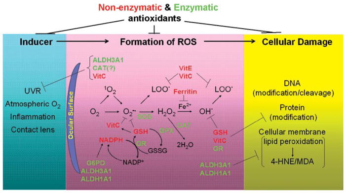
The human eye is subjected constantly to oxidative stress due to daily exposure to sunlight, high metabolic activities, and oxygen tension. Reactive oxygen species generated from environmental insults and pathological conditions render the human eye particularly vulnerable to oxidative damage. The ocular surface composed of the tear film, the cornea, and the aqueous humor forms the first physical and biochemical barrier of the eye and plays a pivotal role in combating free radicals. These ocular compartments are enriched in certain antioxidants in the form of metabolic enzymes or small molecules. Such an antioxidant defense system in the ocular surface is essential for the maintenance of redox homeostasis in the eye and protection against oxidative damage. Herein, we review the properties and functions of key constituent antioxidants of the ocular surface.
Conflict of interest statement
The authors have no commercial or proprietary interest in any concept or product discussed in this article.
Figures


Similar articles
-
Environmental light and endogenous antioxidants as the main determinants of non-cancer ocular diseases.Mutat Res. 2013 Apr-Jun;752(2):153-171. doi: 10.1016/j.mrrev.2013.01.001. Epub 2013 Jan 19. Mutat Res. 2013. PMID: 23337404 Review.
-
The Eye, Oxidative Damage and Polyunsaturated Fatty Acids.Nutrients. 2018 May 24;10(6):668. doi: 10.3390/nu10060668. Nutrients. 2018. PMID: 29795004 Free PMC article. Review.
-
The role of corneal crystallins in the cellular defense mechanisms against oxidative stress.Semin Cell Dev Biol. 2008 Apr;19(2):100-12. doi: 10.1016/j.semcdb.2007.10.004. Epub 2007 Oct 10. Semin Cell Dev Biol. 2008. PMID: 18077195 Review.
-
Oxygen, the Paradox of Life and the Eye.Front Biosci (Landmark Ed). 2024 Sep 13;29(9):319. doi: 10.31083/j.fbl2909319. Front Biosci (Landmark Ed). 2024. PMID: 39344319 Review.
-
Oxidative and antioxidative stress markers in keratoconus: a systematic review and meta-analysis.Acta Ophthalmol. 2021 Sep;99(6):e777-e794. doi: 10.1111/aos.14714. Epub 2020 Dec 23. Acta Ophthalmol. 2021. PMID: 33354927
Cited by
-
Superoxide Dismutase Administration: A Review of Proposed Human Uses.Molecules. 2021 Mar 25;26(7):1844. doi: 10.3390/molecules26071844. Molecules. 2021. PMID: 33805942 Free PMC article.
-
Quantitative analysis of SOD2, ALDH1A1 and MGST1 messenger ribonucleic acid in anterior lens epithelium of patients with pseudoexfoliation syndrome.Mol Vis. 2013 Jun 12;19:1341-9. Print 2013. Mol Vis. 2013. PMID: 23805041 Free PMC article.
-
Emerging roles of oxidative stress in the pathogenesis of pseudoexfoliation syndrome (Review).Exp Ther Med. 2022 Jul 28;24(3):602. doi: 10.3892/etm.2022.11539. eCollection 2022 Sep. Exp Ther Med. 2022. PMID: 35949329 Free PMC article. Review.
-
Method for Collection of Tear Fluid for Evaluation Its Antioxidant Properties.Bull Exp Biol Med. 2023 Aug;175(4):492-496. doi: 10.1007/s10517-023-05893-0. Epub 2023 Sep 28. Bull Exp Biol Med. 2023. PMID: 37768462
-
Effects of collagen cross-linking on the keratoconus metabolic network.Eye (Lond). 2018 Jul;32(7):1271-1281. doi: 10.1038/s41433-018-0075-6. Epub 2018 Mar 26. Eye (Lond). 2018. PMID: 29576618 Free PMC article.
References
-
- Roberts JE. Ocular phototoxicity. J Photochem Photobiol B. 64:136–43. 200. - PubMed
-
- Tenkate TD. Ultraviolet radiation: human exposure and health risks. J Environ Health. 2009;61:9–15.
-
- Andley UP, Rhim JS, Chylack LT, Jr, Fleming TP. Propagation and immortalization of human lens epithelial cells in culture. Invest Ophthalmol Vis Sci. 1994;35:3094–102. - PubMed
-
- Zigman S. Lens UVA photobiology. J Ocul Pharmacol Ther. 2000;16:161–5. - PubMed
Publication types
MeSH terms
Substances
Grants and funding
LinkOut - more resources
Full Text Sources
Other Literature Sources
Medical