

Asymmetric oxidation of giant vesicles triggers curvature-associated shape transition and permeabilization
- PMID: 19948119
- PMCID: PMC2785014
- DOI: 10.1016/j.bpj.2009.08.056
Asymmetric oxidation of giant vesicles triggers curvature-associated shape transition and permeabilization
Abstract
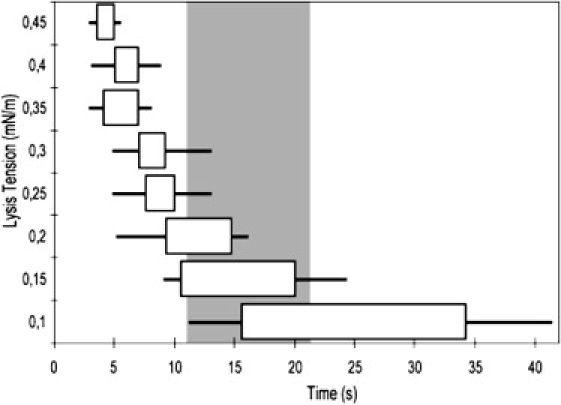
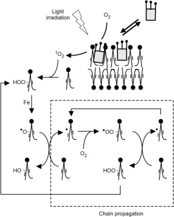
Oxidation of unsaturated lipids is a fundamental process involved in cell bioenergetics as well as in cell death. Using giant unilamellar vesicles and a chlorin photosensitizer, we asymmetrically oxidized the outer or inner monolayers of lipid membranes. We observed different shape transitions such as oblate to prolate and budding, which are typical of membrane curvature modifications. The asymmetry of the shape transitions is in accordance with a lowered effective spontaneous curvature of the leaflet being targeted. We interpret this effect as a decrease in the preferred area of the targeted leaflet compared to the other, due to the secondary products of oxidation (cleaved-lipids). Permeabilization of giant vesicles by light-induced oxidation is observed after a lag and is characterized in relation with the photosensitizer concentration. We interpret permeabilization as the opening of a pore above a critical membrane tension, resulting from the budding of vesicles. The evolution of photosensitized giant vesicle lysis tension was measured and yields an estimation of the effective spontaneous curvature at lysis. Additionally photo-oxidation was shown to be fusogenic.
Figures
Similar articles
-
Lipid Unsaturation Properties Govern the Sensitivity of Membranes to Photoinduced Oxidative Stress.Biophys J. 2019 Mar 5;116(5):910-920. doi: 10.1016/j.bpj.2019.01.033. Epub 2019 Feb 2. Biophys J. 2019. PMID: 30777304 Free PMC article.
-
Curvature-Assisted Vesicle Explosion Under Light-Induced Asymmetric Oxidation.Adv Sci (Weinh). 2024 Oct;11(38):e2400504. doi: 10.1002/advs.202400504. Epub 2024 Aug 13. Adv Sci (Weinh). 2024. PMID: 39136143 Free PMC article.
-
Charged giant unilamellar vesicles prepared by electroformation exhibit nanotubes and transbilayer lipid asymmetry.Sci Rep. 2018 Aug 7;8(1):11838. doi: 10.1038/s41598-018-30286-z. Sci Rep. 2018. PMID: 30087440 Free PMC article.
-
Processes and mechanisms underlying burst of giant unilamellar vesicles induced by antimicrobial peptides and compounds.Biochim Biophys Acta Biomembr. 2024 Jun;1866(5):184330. doi: 10.1016/j.bbamem.2024.184330. Epub 2024 Apr 26. Biochim Biophys Acta Biomembr. 2024. PMID: 38679311 Review.
-
Elementary Processes and Mechanisms of Interactions of Antimicrobial Peptides with Membranes-Single Giant Unilamellar Vesicle Studies.Adv Exp Med Biol. 2019;1117:17-32. doi: 10.1007/978-981-13-3588-4_3. Adv Exp Med Biol. 2019. PMID: 30980351 Review.
Cited by
-
A Molecular Rotor that Measures Dynamic Changes of Lipid Bilayer Viscosity Caused by Oxidative Stress.Chemistry. 2016 Sep 5;22(37):13210-7. doi: 10.1002/chem.201601925. Epub 2016 Aug 3. Chemistry. 2016. PMID: 27487026 Free PMC article.
-
Dynamics of mitochondrial membranes under photo-oxidative stress with high spatiotemporal resolution.Front Cell Dev Biol. 2023 Nov 17;11:1307502. doi: 10.3389/fcell.2023.1307502. eCollection 2023. Front Cell Dev Biol. 2023. PMID: 38046667 Free PMC article.
-
MEMBRANE PROTEIN STRUCTURES AND INTERACTIONS FROM COVALENT LABELING COUPLED WITH MASS SPECTROMETRY.Mass Spectrom Rev. 2022 Jan;41(1):51-69. doi: 10.1002/mas.21667. Epub 2020 Nov 4. Mass Spectrom Rev. 2022. PMID: 33145813 Free PMC article. Review.
-
Thermo-induced vesicular dynamics of membranes containing cholesterol derivatives.Lipids. 2012 Aug;47(8):813-20. doi: 10.1007/s11745-012-3695-9. Epub 2012 Jul 1. Lipids. 2012. PMID: 22752691
-
Formation of Large Hypericin Aggregates in Giant Unilamellar Vesicles-Experiments and Modeling.Biophys J. 2017 Mar 14;112(5):966-975. doi: 10.1016/j.bpj.2017.01.019. Biophys J. 2017. PMID: 28297655 Free PMC article.
References
-
- Girotti A.W. Photodynamic lipid peroxidation in biological systems. Photochem. Photobiol. 1990;51:497–509. - PubMed
-
- Ross R. The pathogenesis of atherosclerosis: a perspective for the 1990s. Nature. 1993;362:801–809. - PubMed
-
- Cadenas E., Parcker L. CRC Press; New York: 1999. Understanding the Process of Aging: the Roles of Mitochondria, Free Radicals and Antioxidants.
-
- Girotti A.W., Kriska T. Role of lipid hydroperoxides in photo-oxidative stress signaling. Antioxid. Redox Signal. 2004;6:301–310. - PubMed
MeSH terms
Substances
LinkOut - more resources
Full Text Sources
