. 2009 Dec 2;97(11):3010-8.
doi: 10.1016/j.bpj.2009.09.017.

Reversible, temperature-dependent supramolecular assembly of aquaporin-4 orthogonal arrays in live cell membranes

Affiliations

Reversible, temperature-dependent supramolecular assembly of aquaporin-4 orthogonal arrays in live cell membranes

Jonathan M Crane et al. Biophys J. .

Abstract

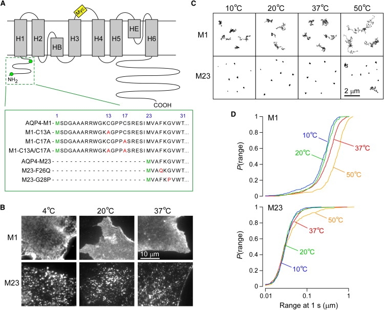
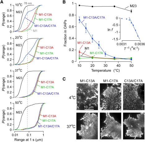
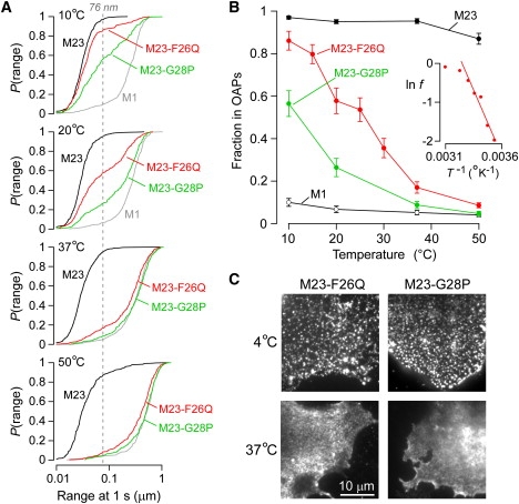
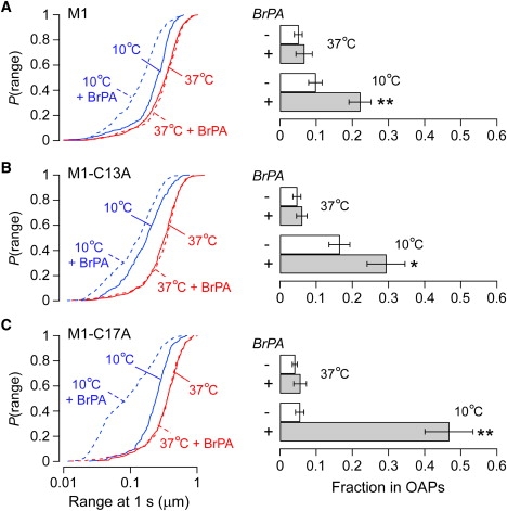
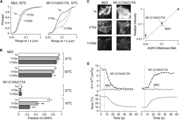
The shorter "M23" isoform of the glial cell water channel aquaporin-4 (AQP4) assembles into orthogonal arrays of particles (OAPs) in cell plasma membranes, whereas the full-length "M1" isoform does not. N-terminal residues are responsible for OAP formation by AQP4-M23 and for blocking of OAP formation in AQP4-M1. In investigating differences in OAP formation by certain N-terminus mutants of AQP4, as measured by freeze-fracture electron microscopy versus live-cell imaging, we discovered reversible, temperature-dependent OAP assembly of certain weakly associating AQP4 mutants. Single-particle tracking of quantum-dot-labeled AQP4 in live cells and total internal reflection fluorescence microscopy showed >80% of M23 in OAPs at 10-50 degrees C compared to <10% of M1. However, OAP formation by N-terminus cysteine-substitution mutants of M1, which probe palmitoylation-regulated OAP assembly, was strongly temperature-dependent, increasing from <10% at 37 degrees C to >70% at 10 degrees C for the double mutant M1-C13A/C17A. OAP assembly by this mutant, but not by native M23, could also be modulated by reducing its membrane density. Exposure of native M1 and single cysteine mutants to 2-bromopalmitate confirmed the presence of regulated OAP assembly by S-palmitoylation. Kinetic studies showed rapid and reversible OAP formation during cooling and OAP disassembly during heating. Our results provide what to our knowledge is the first information on the energetics of AQP4 OAP assembly in plasma membranes.


Figures

Figure 1
Figure 1
Temperature-independent assembly of native AQP4 isoforms M1 and M23. (A) AQP4 schematic showing transmembrane helices (gray), the positions of the inserted Myc sequence (yellow) in the second extracellular loop, and Met1 and Met23 (green) in the cytoplasmic N-terminal domain. N-terminus sequences of the AQP4 mutants used in this study are shown in the expanded green box. (B) TIRF micrographs of Alexa-labeled COS-7 cells transfected with M1 (upper) or M23 (lower) and fixed at the indicated temperature. Scale bar, 10 μm. (C) Representative trajectories from AQP4 isoforms M1 (upper) and M23 (lower) diffusing in the membrane of live COS-7 cells at the indicated temperatures. Scale bar, 2 μm. (D) Cumulative probability distribution of ranges at 1 s (P(range)) for AQP4 isoforms M1 (upper) and M23 (lower) recorded at 10°C (blue), 20°C (green), 37°C (red), and 50°C (orange).
Figure 2
Figure 2
Temperature-dependent OAP assembly of a double cysteine mutant of AQP4-M1. (A) P(range) for single cysteine mutants M1-C13A (red) and M1-C17A (green), and double cysteine mutant M1-C13A/C17A (blue) at indicated temperatures. P(range) for M23 (black) and M1 (gray) are shown for reference. Dashed line indicates the 95th percentile of the range of M23 at 20°C. (B) Estimated fraction of M23 (black), M1 (white), M1-C13A (red), M1-C17A (green), and M1-C13A/C17A (blue) forming OAPs versus temperature, based on the 95th percentile of the range for M23 at 20°C. Error bars represent the mean ± SE. (Inset) Natural log of the fraction of free M1-C13A/C17A tetramers plotted against the inverse of absolute temperature. (C) TIRF micrographs of Alexa-labeled COS-7 cells transfected with M1-C13A (left), M1-C17A (middle), or M1-C13A/C17A (right) and fixed at 4°C (upper) or 37°C (lower). Scale bar, 10 μm.
Figure 3
Figure 3
Temperature-dependent OAP assembly of weakly associating mutants of AQP4-M23. (A) P(range) for point mutants M23-F26Q (red) and M23-G28P (green) at the indicated temperature. P(range) for M23 (black) and M1 (gray) are shown for comparison. Dashed line indicates 95th percentile of the range of M23 at 20°C. (B) Estimated fraction of M23 (black), M1 (white), M23-F26Q (red), and M23-G28P (green) forming OAPs as a function of temperature. Error bars represent the mean ± SE. (Inset) Natural log of the fraction of free M23-F26Q tetramers plotted against the inverse of absolute temperature. (C) TIRF micrographs of Alexa-labeled COS-7 cells transfected with M23-F26Q (left) or M23-G28P (right) and fixed at 4°C (upper) or 37°C (lower). Scale bar, 10 μm.
Figure 4
Figure 4
S-palmitoylation affects temperature-dependent assembly of AQP4-M1. (A) (Left) P(range) of native M1 at 37°C (red) and 10°C (blue) under control conditions (solid line) and after overnight incubation with 2-bromopalmitate (dashed line). (Right) Estimated fractions of AQP4 tetramers in OAPs with or without 2-bromopalmitate. P < 0.05; ∗∗P < 0.01. (B) The same experiment as in A, but with single cysteine mutant M1-C13A. (C) The same experiment as in A, but with single cysteine mutant M1-C17A. Error bars represent the mean ± SE.
Figure 5
Figure 5
Membrane density-dependence and kinetics of assembly of AQP4 in OAPs. (A) P(range) of native M23 at 50°C (left) and M1-C13A/C17A at 10°C (right) without (solid line) and with (dashed line) dilution of the AQP4-encoding plasmid DNA by 10× (dashed line) or 100× (dotted line) with empty plasmid. (B) Estimated fraction of M23 (upper) and M1-C13A/C17A (lower) assembled in OAPs at indicated temperature and relative AQP4 concentration. P < 0.05; ∗∗P < 0.01. Error bars represent the mean ± SE. (C) (Left) Fluorescence micrographs after surface labeling of COS-7 cells with Alexa Fluor 488 (intensity enhanced to visualize cells at 100× dilution). (Right) Average relative intensities versus AQP4 dilution ratios for M23 (open circles) and M1-C13A/C17A (solid circles). Error bars represent the mean ± SE. (D) Average diffusion coefficient (upper) of M23 (open circles) and M1-C13A/C17A (solid circles), and solution temperature (lower), versus time during rapid temperature drop or rise. Dashed lines indicate the time of switching between warm and cold perfusates.

References

    1. Hasegawa H., Ma T., Skach W., Matthay M.A., Verkman A.S. Molecular cloning of a mercurial-insensitive water channel expressed in selected water-transporting tissues. J. Biol. Chem. 1994;269:5497–5500. - PubMed
    1. Jung J.S., Bhat R.V., Preston G.M., Guggino W.B., Baraban J.M. Molecular characterization of an aquaporin cDNA from brain: candidate osmoreceptor and regulator of water balance. Proc. Natl. Acad. Sci. USA. 1994;91:13052–13056. - PMC - PubMed
    1. Frigeri A., Gropper M.A., Turck C.W., Verkman A.S. Immunolocalization of the mercurial-insensitive water channel and glycerol intrinsic protein in epithelial cell plasma membranes. Proc. Natl. Acad. Sci. USA. 1995;92:4328–4331. - PMC - PubMed
    1. Verkman A.S., Binder D.K., Bloch O., Auguste K., Papadopoulos M.C. Three distinct roles of aquaporin-4 in brain function revealed by knockout mice. Biochim. Biophys. Acta. 2006;1758:1085–1093. - PubMed
    1. Hiroaki Y., Tani K., Kamegawa A., Gyobu N., Nishikawa K. Implications of the aquaporin-4 structure on array formation and cell adhesion. J. Mol. Biol. 2006;355:628–639. - PubMed

Publication types

MeSH terms

LinkOut - more resources