

Beta frequency synchronization in basal ganglia output during rest and walk in a hemiparkinsonian rat
- PMID: 19948166
- PMCID: PMC3384738
- DOI: 10.1016/j.expneurol.2009.11.016
Beta frequency synchronization in basal ganglia output during rest and walk in a hemiparkinsonian rat
Abstract
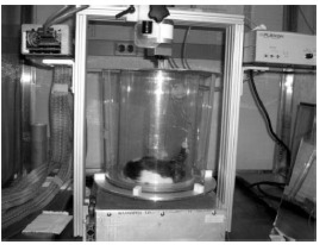
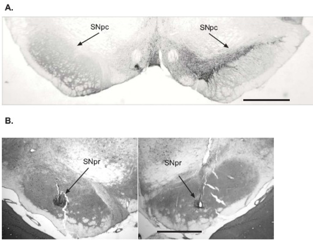
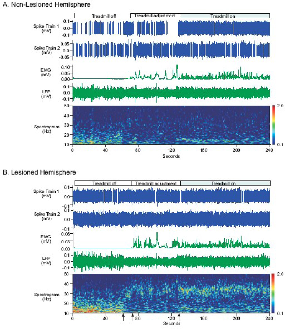
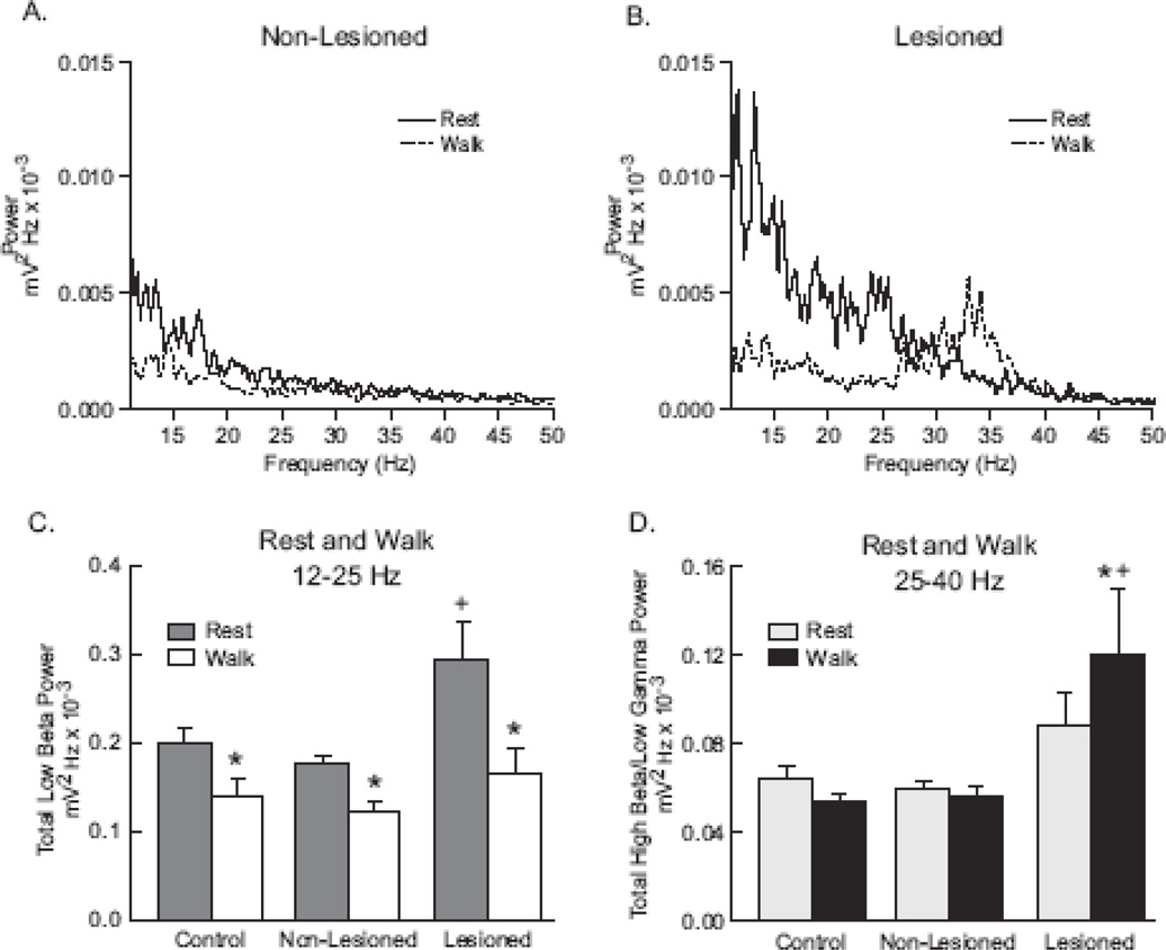
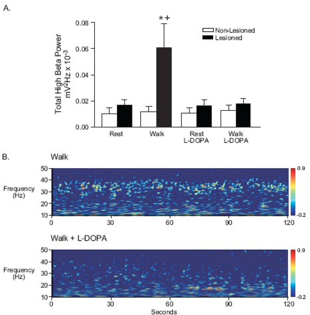
Synchronized oscillatory neuronal activity in the beta frequency range has been observed in the basal ganglia of Parkinson's disease patients and hypothesized to be antikinetic. The unilaterally lesioned rat model of Parkinson's disease allows examination of this hypothesis by direct comparison of beta activity in basal ganglia output in non-lesioned and dopamine cell lesioned hemispheres during motor activity. Bilateral substantia nigra pars reticulata (SNpr) recordings of units and local field potentials (LFP) were obtained with EMG activity from the scapularis muscle in control and unilaterally nigrostriatal lesioned rats trained to walk on a rotary treadmill. After left hemispheric lesion, rats had difficulty walking contraversive on the treadmill but could walk in the ipsiversive direction. During inattentive rest, SNpr LFP power in the 12-25 Hz range (low beta) was significantly greater in the dopamine-depleted hemisphere than in non-lesioned and control hemispheres. During walking, low beta power was reduced in all hemispheres, while 25-40 Hz (high beta) activity was selectively increased in the lesioned hemisphere. High beta power increases were reduced by l-DOPA administration. SNpr spiking was significantly more synchronized with SNpr low beta LFP oscillations during rest and high beta LFP oscillations during walking in the dopamine-depleted hemispheres compared with non-lesioned hemispheres. Data show that dopamine loss is associated with opposing changes in low and high beta range SNpr activity during rest and walk and suggest that increased synchronization of high beta activity in SNpr output from the lesioned hemisphere during walking may contribute to gait impairment in the hemiparkinsonian rat.
Published by Elsevier Inc.
Figures

References
-
- Albin RL, Young AB, Penney JB. The functional anatomy of basal ganglia disorders. Trends Neurosci. 1989;12:366–375. - PubMed
-
- Alegre M, Alonso-Frech F, Rodriguez-Oroz M, Guridi J, Zamarbide I, Valencia M, Manrique M, Obeso J, Artieda J. Movement-related changes in oscillatory activity in the human nucleus: ipsilateral vs. contralateral movements. Eur. J. Neurosci. 2005;22:2315–2324. - PubMed
-
- Alexander GE, Crutcher MD. Functional architecture of basal ganglia circuits: neural substrates of parallel processing. Trends Neurosci. 1990;13:266–271. - PubMed
-
- Alonso-Frech F, Zamarbide I, Alegre M, Rodriguez-Oroz M, Guridi J, Manrique M, Valencia M, Artieda J, Obeso J. Slow oscillatory activity and levodopa-induced dyskinesias in Parkinson's disease. Brain. 2006;129:1748–1757. - PubMed
-
- Anderson M, Yoshida M. Electrophysiological evidence for branching nigral projections to thalamus and superior colliculus. Brain Res. 1977;137:361–364. - PubMed
Publication types
MeSH terms
Substances
Grants and funding
LinkOut - more resources
Full Text Sources
Other Literature Sources
Medical
Miscellaneous
