

Depth of submucosal invasion does not predict lymph node metastasis and survival of patients with esophageal carcinoma
- PMID: 19948247
- PMCID: PMC2834861
- DOI: 10.1016/j.cgh.2009.11.016
Depth of submucosal invasion does not predict lymph node metastasis and survival of patients with esophageal carcinoma
Abstract
Background & aims: There is controversy over the outcomes of esophageal adenocarcinoma with superficial submucosal invasion. We evaluated the impact of depth of submucosal invasion on the presence of metastatic lymphadenopathy and survival in patients with esophageal adenocarcinoma.
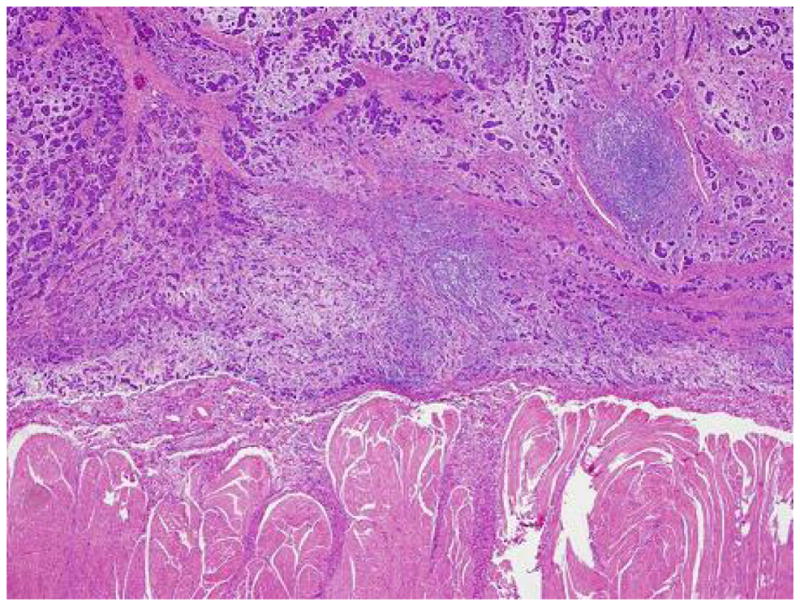
Methods: Pathology reports of esophagectomy samples collected from 1997 to 2007 were reviewed. Specimens from patients with esophageal adenocarcinoma and submucosal invasion were reviewed and classified as superficial (upper 1 third, sm1) or deep (middle third, sm2 or deepest third, sm3) invasion. Outcomes studied were presence of metastatic lymphadenopathy and overall survival. Variables of interest were analyzed as factors that affect overall and cancer-free survival using Cox proportional hazards modeling. A multivariate model was constructed to establish independent associations with survival.
Results: The study included 80 patients; 31 (39%) had sm1 carcinoma, 23 (29%) had sm2 carcinoma, and 26 (33%) had sm3 carcinoma. Superficial and deep submucosal invasion were associated with substantial rates of metastatic lymphadenopathy (12.9% and 20.4%, respectively). The mean follow-up time was 40.5 +/- 4 months and the mean overall unadjusted survival time was 53.8 +/- 4.1 months. Factors significantly associated with reduced survival time included the presence of metastatic lymph nodes (hazard ratio [HR], 2.89; confidence interval [CI], 1.13-6.88) and esophageal cancer recurrence (HR 6.39, CI 2.40-16.14), but not depth of submucosal invasion.
Conclusions: Patients with sm1 esophageal carcinoma have substantial rates of metastatic lymphadenopathy. Endoscopic treatment of superficial submucosal adenocarcinoma is not advised for patients that are candidates for surgery.
Copyright 2010 AGA Institute. Published by Elsevier Inc. All rights reserved.
Figures


Comment in
-
Early adenocarcinoma of the esophagus - clinical relevance of depth of infiltration into the submucosa.Clin Gastroenterol Hepatol. 2011 Mar;9(3):277; author reply 277-8. doi: 10.1016/j.cgh.2010.10.004. Epub 2010 Oct 15. Clin Gastroenterol Hepatol. 2011. PMID: 20951839 No abstract available.
References
-
- Falk GW. Barrett’s esophagus. Gastroenterology. 2002;122:1569–91. - PubMed
-
- Devesa SS, Blot WJ, Fraumeni JF., Jr Changing patterns in the incidence of esophageal and gastric carcinoma in the United States. Cancer. 1998;83:2049–53. - PubMed
-
- van Lanschot JJ, Hulscher JB, Buskens CJ, Tilanus HW, ten Kate FJ, Obertop H. Hospital volume and hospital mortality for esophagectomy. Cancer. 2001;91:1574–8. - PubMed
-
- Pacifico RJ, Wang KK, Wongkeesong LM, Buttar NS, Lutzke LS. Combined endoscopic mucosal resection and photodynamic therapy versus esophagectomy for management of early adenocarcinoma in Barrett’s esophagus. Clin Gastroenterol Hepatol. 2003;1:252–7. - PubMed
-
- Buttar NS, Wang KK, Lutzke LS, Krishnadath KK, Anderson MA. Combined endoscopic mucosal resection and photodynamic therapy for esophageal neoplasia within Barrett’s esophagus. Gastrointest Endosc. 2001;54:682–8. - PubMed
Publication types
MeSH terms
Grants and funding
LinkOut - more resources
Full Text Sources
Medical
