. 2010 Jan;10(1):9-15.
doi: 10.1016/j.gep.2009.11.005. Epub 2009 Dec 3.

Autism susceptibility candidate 2 (Auts2) encodes a nuclear protein expressed in developing brain regions implicated in autism neuropathology

Affiliations

Autism susceptibility candidate 2 (Auts2) encodes a nuclear protein expressed in developing brain regions implicated in autism neuropathology

Francesco Bedogni et al. Gene Expr Patterns. 2010 Jan.

Abstract

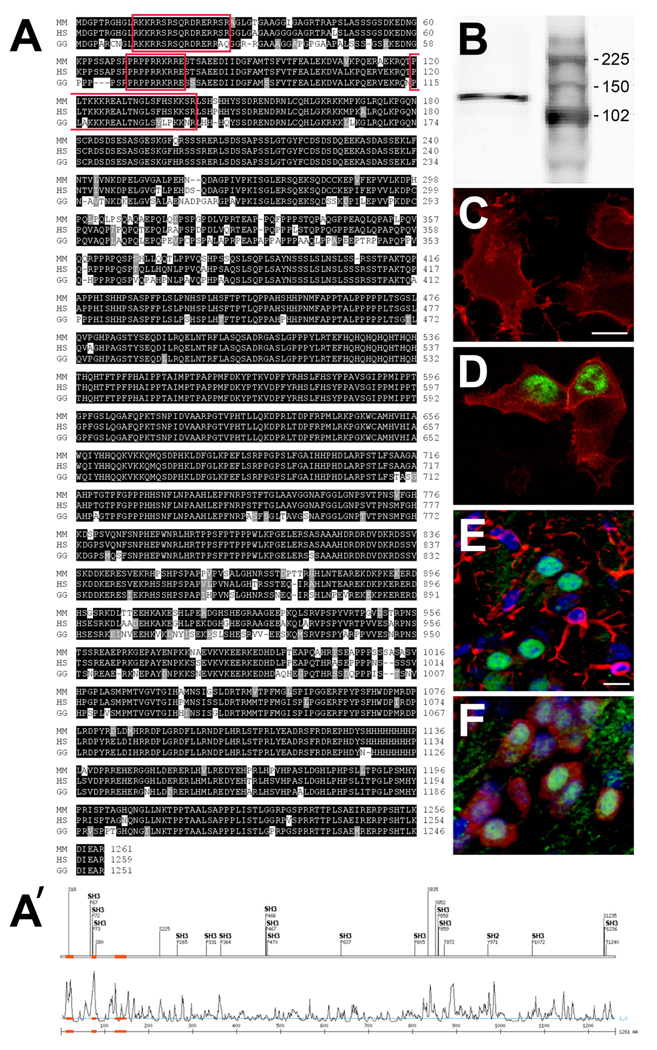
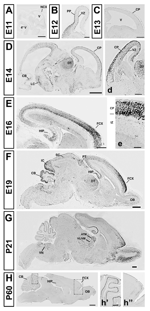
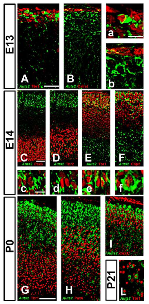
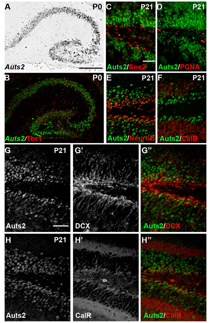
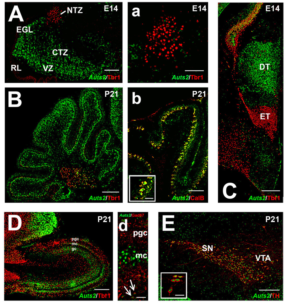
Autism susceptibility candidate 2 (Auts2) is a gene associated with autism and mental retardation, whose function is unknown. Expression of Auts2 mRNA and protein were studied in the developing mouse brain by in situ hybridization, immunohistochemistry, and western blotting. Auts2 mRNA was highly expressed in the developing cerebral cortex and cerebellum, regions often affected by neuropathological changes in autism, and a few other brain regions. On embryonic day (E) 12, Auts2 mRNA was expressed in the cortical preplate, where it colocalized with Tbr1, a transcription factor specific for postmitotic projection neurons. From E16 to postnatal day 21, Auts2 was expressed most abundantly in frontal cortex, hippocampus and cerebellum, including Purkinje cells and deep nuclei. High levels of Auts2 were also detected in developing dorsal thalamus, olfactory bulb, inferior colliculus and substantia nigra. Auts2 protein showed similar regional expression patterns as the mRNA. At the cellular level, Auts2 protein was localized in the nuclei of neurons and some neuronal progenitors.


Figures

Figure 1
Figure 1
Analysis of Auts2 protein sequence and cellular localization. A: alignment of Auts2 amino acid sequence from Mus Musculus (MM), Homo Sapiens (HS) and Gallus Gallus (GG). Conserved residues are represented in black, similar residues in gray, and differently charged residues are not marked. Nuclear localization sequences (NLS) are highlighted in red. A’: Upper trace shows putative SH2 and SH3 domains. Lower trace shows surface accessibility plot with putative NLS (red). B: The specificity of Auts2 antibody was tested by Western blotting on E14 protein mouse cortical extract, revealing a band at 139.8 kDa. C–D: Auts2 protein was evaluated by transfection of membrane tomato (red) alone (C) or with Auts2 (green, D) in 293T cells. Auts2 was detected in the nuclei of cells transfected with the Auts2 expression plasmid. E–F: Expression of Auts2 (green) in postnatal day (P) 21 cortex showed no obvious colocalization with GFAP (red, E). However, Auts2 was detected in neurons, as shown by colocalization with NeuN (red, P21, F). Scale bars: C–D–E–F: 10 µm.
Figure 2
Figure 2
Auts2 mRNA expression patterns at different embryonic and postnatal ages revealed by in situ hybridization (ISH). NCX: neocortex; V: ventricle; PP: preplate; VZ: ventricular zone; CP: cortical plate; DT: dorsal thalamus; CB: cerebellum; LC: locus ceruleus; OB: olfactory bulbs; FCX: frontal cortex; HIP: hippocampus; PT: pretectum; SC: superior colliculus; IC: inferior colliculus; ATN: anterior thalamic nuclei; VL/VM: ventrolateral/ventromedial nuclei; VN: vestibular nuclei. Scale bars: A: 500 µm; B: 200 µm; C–D–F–G: 1mm; d: 100 µm; E: 200 µm, e: 75 µm, H: 500 µm, h’ (for h’–h”) 100 µm. Ages: A: E11, B: E12, C: E13, D: E14, E: E16, F: E19, G: P21.
Figure 3
Figure 3
Auts2 mRNA expression in the developing cortex, revealed by ISH (green, pseudocolor). The pattern of expression is studied in association with other markers detected by immunohistochemistry (red) in the same section. A, a, E, e, G: Tbr1; B, b: calretinin; C, c: Pax6; D, d: Tbr2; F, f, H: Ctip2; I: Cux1. CP: cortical plate; IZ: intermediate zone. Scale bars: A: 50 µm (applies to A, B); a: 20 µm (applies to a, b); C: 50 µm (applies to C–F); c: 20 µm (applies to c–f); G: 100 µm (applies to G–I). Ages: A–B, a–b: E13, C–F, c–f: E14, G–I, g–i: P0.
Figure 4
Figure 4
Auts2 mRNA and protein expression in the developing hippocampus. A–B: At P0, Auts2 expression, as detected by ISH (gray or green), was apparent throughout the developing hippocampus where it colocalized with Tbr1 immunofluorescence (red). C–H: Auts2 protein, detected by immunofluorescence at P21, was apparent throughout the granule cell layer (GCL) of the dentate gyrus. Coexpression with specific markers of progenitor cells and differentiating neurons was assessed by double immunofluorescent labeling with C) Sox2; D) PCNA; E) NeuroD1; F) calbindin (CalB); G–G”) DCX; H–H”) calretinin (CalR). Scale bars: A 100 µm (applies to A, B); C: 50 µm (applies to C–F) G: 50 µm (applies to G–H”).
Figure 5
Figure 5
Expression of Auts2 mRNA in brain regions outside the cortex at different stages of development. A–a) Double labeling for Auts2 mRNA revealed by in situ hybridization (green) and Tbr1 protein by immunofluorescence (red) in the E14 cerebellum. NTZ: nuclear transitory zone; EGL: external granular layer; CTZ: cortical transitory zone; RL: rhombic lip; VZ: ventricular zone. B) Postnatal day (P) 21 cerebellum labeled with Auts2 (green) in relationship to Tbr1 (red); b) expression of Auts2 protein (green, immunofluorescence) in Purkinje cells identified by calbindin immunofluorescence (red). C) Auts2 mRNA (green) was expressed in the dorsal thalamus (DT), in a complementary fashion compared to the eminentia thalami (ET), stained by Tbr1 (red, immunofluorescence). D) Auts2 mRNA (green) was localized in the P21 olfactory bulb mitral cells (MC) and the periglomerular cells (PGC); overlap with Tbr1 (red) was found in some MC. Also, some overlap of Auts2 mRNA (green) with GAD67 (red, d), a marker of GABAergic neurons, was detected in the GC (arrows). E) Strong coexpression of Auts2 protein (green) and TH (red) was found in the substantia nigra (SN) and the ventral tegmental area (VTA). Scale bars: A: 100 µm; a: 50 µm; B: 100 µm; b: 50 µm; inset b: 20 µm; C: 200 µm; D: 100 µm; d: 30 µm; E: 100 µm; inset E: 20 µm. Ages: A–a: E14, B–b: P21, C: E14, D–d: P21, E: P21.

Similar articles

Cited by

References

    1. Amaral D, Schumann C, Nordahl C. Neuroanatomy of autism. Trends. Neurosci. 2008;31:137–145. - PubMed
    1. Arlotta P, Molyneaux BJ, Chen J, Inoue J, Kominami R, Macklis JD. Neuronal subtype-specific genes that control corticospinal motor neuron development in vivo. Neuron. 2005;45:207–221. - PubMed
    1. Autism Genome Project Consortium. Mapping autism risk loci using genetic linkage and chromosomal rearrangements. Nat. Genet. 2007;39(3):319–328. - PMC - PubMed
    1. Ballas N, Lioy D, Grunseich C, Mandel G. Non–cell autonomous influence of MeCP2-deficient glia on neuronal dendritic morphology. Nat. Neurosci. 2009;12:311–317. - PMC - PubMed
    1. Bauman ML, Kemper TL. Neuroanatomic observations of the brain in autism: a review and future directions. Int. J. Dev. Neurosci. 2005;23(2–3):183–187. - PubMed

Publication types