

Correlation of cutaneous tension distribution and tissue oxygenation with acute external tissue expansion
- PMID: 19948443
- PMCID: PMC3352288
- DOI: 10.1186/2047-783x-14-11-280
Correlation of cutaneous tension distribution and tissue oxygenation with acute external tissue expansion
Abstract
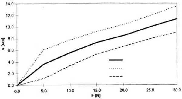
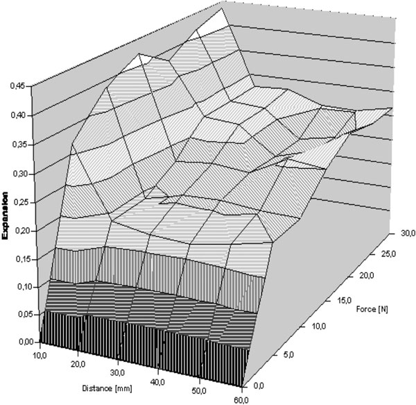
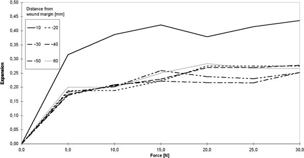
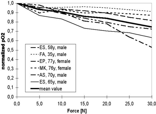
Today, the biomechanical fundamentals of skin expansion are based on viscoelastic models of the skin. Although many studies have been conducted in vitro, analyses performed in vivo are rare. Here, we present in vivo measurements of the expansion at the skin surface as well as measurement of the corresponding intracutaneous oxygen partial pressure. In our study the average skin stretching was 24%, with a standard deviation of 11%, excluding age or gender dependency. The measurement of intracutaneous oxygen partial pressure produced strong inter-individual fluctuations, including initial values at the beginning of the measurement, as well as varying individual patient reactions to expansion of the skin. Taken together, we propose that even large defect wounds can be closed successfully using the mass displacement caused by expansion especially in areas where soft, voluminous tissue layers are present.
Figures
References
-
- Greenbaum SS. Intraoperative tissue expansion with the Foley catheter. J Dermatol Surg Oncol. 1993;19(12):1079–83. - PubMed
-
- Frechet P. Scalp extension. J Dermatol Surg Oncol. 1993;19(7):616–22. - PubMed
-
- Ilizarov GA. The tension-stress effect on the genesis and growth of tissues. Part I. The influence of stability of fixation and soft-tissue preservation. Clin Orthop Relat Res. 1989. pp. 249–81. - PubMed
-
- Ilizarov GA. The tension-stress effect on the genesis and growth of tissues: Part II. The influence of the rate and frequency of distraction. Clin Orthop Relat Res. 1989. pp. 263–85. - PubMed
-
- Vandenburgh HH. Mechanical forces and their second messengers in stimulating cell growth in vitro. Am J Physiol. 1992;262(3 Pt 2):R350–5. - PubMed
MeSH terms
Substances
LinkOut - more resources
Full Text Sources
