Review
. 2009 Nov 2;187(3):319-26.
doi: 10.1083/jcb.200908074.

The ubiquitin landscape at DNA double-strand breaks

Affiliations
Review

The ubiquitin landscape at DNA double-strand breaks

Troy E Messick et al. J Cell Biol. .

Abstract

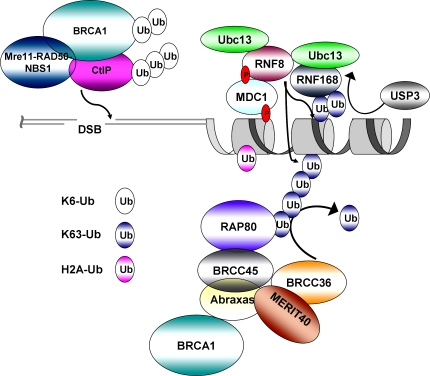
The intimate relationship between DNA double-strand break (DSB) repair and cancer susceptibility has sparked profound interest in how transactions on DNA and chromatin surrounding DNA damage influence genome integrity. Recent evidence implicates a substantial commitment of the cellular DNA damage response machinery to the synthesis, recognition, and hydrolysis of ubiquitin chains at DNA damage sites. In this review, we propose that, in order to accommodate parallel processes involved in DSB repair and checkpoint signaling, DSB-associated ubiquitin structures must be nonuniform, using different linkages for distinct functional outputs. We highlight recent advances in the study of nondegradative ubiquitin signaling at DSBs, and discuss how recognition of different ubiquitin structures may influence DNA damage responses.


Figures

Figure 1.
Figure 1.
Structural topology of ubiquitin chains. (A) Surface representation of ubiquitin. All seven lysines (K6, K11, K27, K29, K33, K48, and K63) and the amino-terminus (M1), shown in blue, can be conjugated to the carboxy terminus of another ubiquitin molecule. The hydrophobic patch (L8, I44, and V70), shown in green, is recognized by several ubiquitin-binding proteins (PDB accession no. 1UBQ; Vijay-Kumar, et al., 1987). (B) Model of K63-tetraubiquitin (based on K63-diubiquitin, PDB accession no. 3A1Q; Sato et al., 2009). K63-tetraubiquitin forms long chains, exposing the I44 hydrophobic patch in green, to ubiquitin-binding proteins. (C) Model of K48-tetraubiquitin (based on K48-diubiquitin, PDB accession no. 1F9J; Phillips et al., 2001). K48-tetraubiquitin forms a compact structure, where the I44 hydrophobic patch in green is largely buried.
Figure 2.
Figure 2.
Structural basis for the specificity of RAP80 binding to ubiquitin. (A) RAP80 UIM1 and UIM2 bound to K63-diubiquitin. The UIM domains of RAP80 (magenta) recognize the I44 hydrophobic patches (green) of ubiquitin. The inter-UIM region (pink) adopts an α-helical fold. (Sato et al., 2009) (B) Comparison of the RAP80 bound and unbound forms of K63-linked diubiquitin. The distal ubiquitin was superimposed to show differences in the proximal ubiquitin between bound (yellow) and unbound (pale blue) of K63-linked diubiquitin. RAP80 induces an ∼45° rotation about the carboxy-terminal region glycine–glycine axis of K63-linked diubiquitin (Komander et al., 2009; Sato et al., 2009).
Figure 3.
Figure 3.
Model for differential ubiquitin-related recognition and repair activities at DSBs. Ubiquitin chains of differing topologies, as indicated, covalently linked to different substrate proteins create a varied ubiquitin landscape at DSBs. Differential recognition of this ubiquitin environment by DNA repair proteins targets repair and checkpoint activities to the appropriate location adjacent to DSBs.

References

    1. Al-Hakim A.K., Zagorska A., Chapman L., Deak M., Peggie M., Alessi D.R. 2008. Control of AMPK-related kinases by USP9X and atypical Lys(29)/Lys(33)-linked polyubiquitin chains. Biochem. J. 411:249–260 10.1042/BJ20080067 - DOI - PubMed
    1. Andreassen P.R., D’Andrea A.D., Taniguchi T. 2004. ATR couples FANCD2 monoubiquitination to the DNA-damage response. Genes Dev. 18:1958–1963 10.1101/gad.1196104 - DOI - PMC - PubMed
    1. Bekker-Jensen S., Lukas C., Kitagawa R., Melander F., Kastan M.B., Bartek J., Lukas J. 2006. Spatial organization of the mammalian genome surveillance machinery in response to DNA strand breaks. J. Cell Biol. 173:195–206 10.1083/jcb.200510130 - DOI - PMC - PubMed
    1. Berkovich E., Monnat R.J., Jr., Kastan M.B. 2007. Roles of ATM and NBS1 in chromatin structure modulation and DNA double-strand break repair. Nat. Cell Biol. 9:683–690 10.1038/ncb1599 - DOI - PubMed
    1. Botuyan M.V., Lee J., Ward I.M., Kim J.E., Thompson J.R., Chen J., Mer G. 2006. Structural basis for the methylation state-specific recognition of histone H4-K20 by 53BP1 and Crb2 in DNA repair. Cell. 127:1361–1373 10.1016/j.cell.2006.10.043 - DOI - PMC - PubMed

Publication types