. 2009 Nov 16;187(4):463-71.
doi: 10.1083/jcb.200906161. Epub 2009 Nov 9.

A dual function for Pex3p in peroxisome formation and inheritance

Affiliations

A dual function for Pex3p in peroxisome formation and inheritance

Joanne M Munck et al. J Cell Biol. .

Abstract

Saccharomyces cerevisiae Pex3p has been shown to act at the ER during de novo peroxisome formation. However, its steady state is at the peroxisomal membrane, where its role is debated. Here we show that Pex3p has a dual function: one in peroxisome formation and one in peroxisome segregation. We show that the peroxisome retention factor Inp1p interacts physically with Pex3p in vitro and in vivo, and split-GFP analysis shows that the site of interaction is the peroxisomal membrane. Furthermore, we have generated PEX3 alleles that support peroxisome formation but fail to support recruitment of Inp1p to peroxisomes, and as a consequence are affected in peroxisome segregation. We conclude that Pex3p functions as an anchor for Inp1p at the peroxisomal membrane, and that this function is independent of its role at the ER in peroxisome biogenesis.


Figures

Figure 1.
Figure 1.
Isolation of a new class of pex3 mutants. (A) pex3Δ cells expressing the lumenal peroxisomal marker GFP-PTS1 were transformed with plasmids containing a WT PEX3 allele or a representative member of each pex3 mutant class. inp1Δ cells are included for comparison. (B) Quantitative description of peroxisome distribution in WT, pex3Δ, inp1Δ, inp2Δ, and the class III mutants pex3-1 and pex3-2. Overnight cultures were diluted and grown for 6 h in selective glucose medium, examined by epifluorescence and phase contrast, and scored for peroxisome distribution. More than 100 budding cells were analyzed for each strain. Three independent experiments were performed. Error bars represent SEM. (C) pex3-1 cells expressing GFP-PTS1 were spotted on an agarose pad and peroxisome distribution was followed with time. A and C show merged brightfield (blue) and fluorescent images (green). Bar, 5 µm.
Figure 2.
Figure 2.
Inp1p localization in peroxisome-deficient cells. A plasmid that expresses Inp1p-GFP under the control of its endogenous promoter was transformed into WT (A), pex19Δ (A–C), and pex3Δ (A) cells expressing HcRed-PTS1 (A), Sec66p-HcRed (B), and Pex3p-mRFP (C). The strains were grown on selective medium and examined by epifluorescence and phase contrast. For A and C, multiple epifluorescence images were acquired in the z axis and flattened into a single image. For B, a single focal plane was taken. Bar, 5 µm.
Figure 3.
Figure 3.
Inp1p binds directly to the cytosolic domain of Pex3p in vitro. GST-Pex3p (40–441) and GST were bound to glutathione Sepharose beads and incubated with a detergent lysate of spheroplasts expressing HA-tagged Inp1p and Mvp1p at endogenous levels (A). After extensive washing, the bound fraction and lysate were analyzed by SDS-PAGE and immunoblotting using the HA monoclonal 12CA5. Yeast lysates (YL) represent 5% of the lysate added to the beads and analyzed by blotting. Because the signal of Inp1p-HA was too low in the YL, 5 times more lysate was reloaded on a separate gel and compared with the GST- and GST-Pex3–bound fraction (right-hand panel). Bottom panel shows Coomassie staining. (B) GST-Inp1p and GST were bound to glutathione Sepharose and incubated with a lysate of E. coli expressing either 6xHIS-tagged Pex3p (40–441) or HIS-tag only, or with lysis buffer only (−). After extensive washing, bound fractions were analyzed by SDS-PAGE and Coomassie staining of the gel. A lane was included with partially purified 6xHIS-Pex3p as control. M, molecular weight marker. Arrow indicates 6xHIS-Pex3p. Asterisks indicate multiple GST-Inp1p fragments. EL, E. coli lysate of 6xHIS-Pex3p–expressing cells.
Figure 4.
Figure 4.
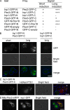
Inp1p interacts with cytosolic domain of Pex3p in vivo. (A–C) Split-GFP analysis between Inp1p and Pex3p in WT cells. Tagged proteins were expressed under control of the GAL1 promoter for 4 h (short) or 8 h (long) and scored for the presence and intensity of fluorescence (A). −, no signal; +, faint; +++, strong. (B) Selected images of WT cells induced for 4 or 8 h. (C) WT cells were induced to express Inp1p-GFP-N and Pex3p-GFP-C for 4 h, followed by mating with pex3Δ cells expressing HcRed-PTS1, and imaging 2 h after mating. GFP and HcRed signals overlap in mated cell (arrow). (D) The expression of a chimeric protein consisting of the cytosolic domain of Pex3p fused at its N terminus to Tom70p and tagged at its C terminus with mRFP (mito-Pex3p-mRFP) was induced on galactose for 3 h in pex3Δ cells expressing Inp1p-GFP under control of its endogenous promoter. Image shows two budding cells, one of which is expressing mito-Pex3p-RFP that recruits Inp1p-GFP. The strains were grown on selective medium and examined by epifluorescence and phase contrast. Multiple epifluorescence images were acquired in the z axis and flattened into a single image. The brightfield image is blue in the merged pictures. Bar, 5 µm.
Figure 5.
Figure 5.
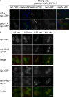
Pex3-1p fails to recruit Inp1p-GFP to peroxisomes. (A) WT and pex3-1 cells expressing Inp1p-GFP at endogenous levels were mated with pex3Δ cells expressing HcRed-PTS1 and imaged after 2 h. (B) The cytosolic domain of Pex3p and Pex3-1p was redirected to mitochondria by fusion to Tom70p in pex3Δ cells expressing Inp1p-GFP at endogenous levels. Mito-Pex3-mRFP and mito-Pex3-1-mRFP expression was induced on galactose medium for times indicated. Signals of mito-Pex3p and mito-Pex3-1p are directly comparable; Inp1p-GFP signals are more enhanced in pex3-1 cells. The strains were grown on selective medium and examined by epifluorescence and phase contrast. Bar, 5 µm.

Similar articles

Cited by

References

    1. Barnard E., McFerran N.V., Trudgett A., Nelson J., Timson D.J. 2008. Development and implementation of split-GFP-based bimolecular fluorescence complementation (BiFC) assays in yeast. Biochem. Soc. Trans. 36:479–482 10.1042/BST0360479 - DOI - PubMed
    1. Boldogh I.R., Ramcharan S.L., Yang H.C., Pon L.A. 2004. A type V myosin (Myo2p) and a Rab-like G-protein (Ypt11p) are required for retention of newly inherited mitochondria in yeast cells during cell division. Mol. Biol. Cell. 15:3994–4002 10.1091/mbc.E04-01-0053 - DOI - PMC - PubMed
    1. Cerveny K.L., Studer S.L., Jensen R.E., Sesaki H. 2007. Yeast mitochondrial division and distribution require the cortical num1 protein. Dev. Cell. 12:363–375 10.1016/j.devcel.2007.01.017 - DOI - PubMed
    1. Distel B., Erdmann R., Gould S.J., Blobel G., Crane D.I., Cregg J.M., Dodt G., Fujiki Y., Goodman J.M., Just W.W., et al. 1996. A unified nomenclature for peroxisome biogenesis factors. J. Cell Biol. 135:1–3 10.1083/jcb.135.1.1 - DOI - PMC - PubMed
    1. Fagarasanu M., Fagarasanu A., Tam Y.Y., Aitchison J.D., Rachubinski R.A. 2005. Inp1p is a peroxisomal membrane protein required for peroxisome inheritance in Saccharomyces cerevisiae. J. Cell Biol. 169:765–775 10.1083/jcb.200503083 - DOI - PMC - PubMed

Publication types

MeSH terms