. 2009 Nov 16;187(4):513-24.
doi: 10.1083/jcb.200907047. Epub 2009 Nov 9.

Capacity for stochastic self-renewal and differentiation in mammalian spermatogonial stem cells

Affiliations

Capacity for stochastic self-renewal and differentiation in mammalian spermatogonial stem cells

Zhuoru Wu et al. J Cell Biol. .

Abstract

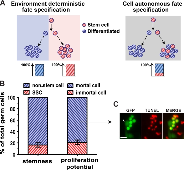
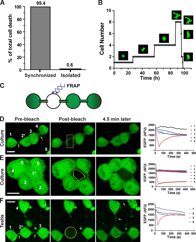
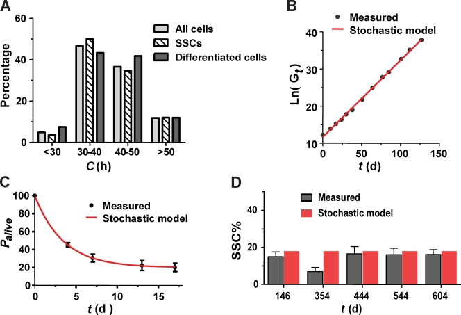
Mammalian spermatogenesis is initiated and sustained by spermatogonial stem cells (SSCs) through self-renewal and differentiation. The basic question of whether SSCs have the potential to specify self-renewal and differentiation in a cell-autonomous manner has yet to be addressed. Here, we show that rat SSCs in ex vivo culture conditions consistently give rise to two distinct types of progeny: new SSCs and differentiating germ cells, even when they have been exposed to virtually identical microenvironments. Quantitative experimental measurements and mathematical modeling indicates that fate decision is stochastic, with constant probability. These results reveal an unexpected ability in a mammalian SSC to specify both self-renewal and differentiation through a self-directed mechanism, and further suggest that this mechanism operates according to stochastic principles. These findings provide an experimental basis for autonomous and stochastic fate choice as an alternative strategy for SSC fate bifurcation, which may also be relevant to other stem cell types.


Figures

Figure 1.
Figure 1.
Persistent heterogeneity of germ cells in stemness and proliferation potential under uniform culture conditions is compatible with a cell-autonomous fate specification model. (A) Environment deterministic and cell-autonomous fate specification models predict homogeneous and heterogeneous composition of SSC progeny, respectively, in a uniform environment. (A, left) In the environment deterministic model, self-renewal and differentiation are controlled by distinct environmental cues (red and blue), which result in homogeneous yet distinct fate outcomes in the red and the blue area. (A, right) In the cell-autonomous fate specification model, occurrence of self-renewal and differentiation does not depend on differential environmental cues, and both SSCs and differentiating progeny can be generated in a uniform environment (gray). (B) Transplantation of germ cells that have been cultured for 20 mo into sterile testes shows that only 16.5 ± 3.9% (mean ± SEM, n = 6) of the population are SSCs, and that 83.5% do not possess stemness. Tracing of sorted single germ cells in culture for 6 mo shows that 79.0 ± 4.1% (mean ± SEM, n = 84 in three indepenent experiments) of the cells perish within a month after transient proliferation, and only 21.0% of the single cells can continue proliferate for at least 6 mo. (C) Dying germ cells show condensed cell bodies and are positive for TUNEL staining. The arrowhead points to a healthy TUNEL-negative germ cell that remains flattened on the culture substrate. Bar, 20 µm.
Figure 2.
Figure 2.
Immortal germ cells are SSCs. (A) Outline of the experimental protocol to yield single cell–derived long-lasting clones and subsequent transplantation of cells from these clones into presterilized recipient rat testes. (B) A wild-type recipient testis transplanted with a long-lasting clone derived from single EGFP-positive germ cell. The donor SSC-derived EGFP-positive spermatogenic colonies (left) indicate the founding cell of the long-lasting clone was an SSC. An enlarged view of individual donor SSC derived colonies (right) showed multiple layers of differentiating germ cells. This testis is representative of the 26 recipient testes, all of which were colonized similarly by donor SSCs from long-lasting clones. White boxes indicate sections that have been enlarged. Bars: (left) 500 µm; (right) 100 µm.
Figure 3.
Figure 3.
Mortal nonstem germ cells in culture are differentiating. (A) Analysis of time-lapse image sequences showed that among germ cells that have divided at least once in culture, 99.4% of the cell deaths (n = 314) are synchronized among sibling groups; only 0.6% died as isolated cells. (B) Images from a time-lapse sequence of a typical mortal germ cell–derived clone (Video 1) and a plot of the cell number within the clone over time show the synchrony of cell divisions and cell death of sibling germ cells in the clone. (C) The experimental scheme for a functional assay specific for intercellular bridges. It involves photobleaching of EGFP within one cell of a group and monitoring of EGFP fluorescence intensity of each cell by time-lapse imaging. (D–F) Time-lapse image sequences before and after photobleaching of the cell indicated by an asterisk, and a time trace of EGFP fluorescence intensity in each cell of a germ cell group in culture (D and E, also see Video 2) or in testis (F). Bleached regions are delineated by yellow lines. Arrows point to visible intercellular bridges. The numbers in D–F indicate which cells correspond to the fluorescence intensity traces shown on the right. Bars, 10 µm.
Figure 4.
Figure 4.
Individual SSCs consistently give rise to SSCs and differentiating germ cells independently of environmental cues. (A) Cultures of single germ cells from two randomly selected long-lasting clones (Nos. 1 and 6) derived from single SSCs showed that they contained 82.7% and 78.4% mortal differentiating cells and 17.3% and 21.6% immortal SSCs, respectively (n = 84). This was similar to their parent cell line (82.7% differentiating and 17.3% SSC, n = 84), which indicates that individual SSCs consistently give rise to both SSCs and differentiating germ cells. (B) Schematic diagram based on the long-term time-lapse imaging of twin daughter cells of a single SSC that gave rise to two clones with different fates. In one clone, all cells died synchronously by day 10, whereas the other continued to live by day 18, which indicates that one of the twin daughter cells differentiated, whereas the other became an SSC. (C and D) Relative positions of the two daughter cells during the time between their birth and next division (Video 3). (C) The solid line indicates the mean diameter (∼13 µm) of the two cells, and black dots mark the distance between the geometric centers of the two cells at the indicated time points. (D) Centroid positions of each daughter cell during the time window are represented by the centers of the red and blue circles, respectively. The diameter of the circles is equal to the mean diameter of the cells to show the area transiently visited by the two cells.
Figure 5.
Figure 5.
A mathematical model for stochastic fate specification describes and predicts germ cell population dynamics in culture. (A) Histogram of cell cycle length in groups of retrospectively identified SSCs (n = 110), differentiating mortal cells (n = 76, synchronized sibling divisions are counted as one), and all cells (n = 186). No significant difference was observed between the SSCs and the differentiating cells. Mean cell cycle length for all cells was 40.0 ± 8.2 h. (B and C) The mathematical model (red line) fits the measured germ cell growth (B, black dots), and the measured germ cell survival curve (C, black dots; mean ± SEM, n = 84 in three independent experiments). (D) The mathematical model predicts a constant SSC content of 17.9% over time (red bars). Dark gray bars indicate the measured SSC content at the indicated time points (mean ± SEM, n > 4).

Similar articles

Cited by

References

    1. Barroca V., Lassalle B., Coureuil M., Louis J.P., Le Page F., Testart J., Allemand I., Riou L., Fouchet P. 2009. Mouse differentiating spermatogonia can generate germinal stem cells in vivo. Nat. Cell Biol. 11:190–196 10.1038/ncb1826 - DOI - PubMed
    1. Bell M.L., Earl J.B., Britt S.G. 2007. Two types of Drosophila R7 photoreceptor cells are arranged randomly: a model for stochastic cell-fate determination. J. Comp. Neurol. 502:75–85 10.1002/cne.21298 - DOI - PubMed
    1. Brawley C., Matunis E. 2004. Regeneration of male germline stem cells by spermatogonial dedifferentiation in vivo. Science. 304:1331–1334 10.1126/science.1097676 - DOI - PubMed
    1. Brinster R.L. 2002. Germline stem cell transplantation and transgenesis. Science. 296:2174–2176 10.1126/science.1071607 - DOI - PMC - PubMed
    1. Brinster R.L., Avarbock M.R. 1994. Germline transmission of donor haplotype following spermatogonial transplantation. Proc. Natl. Acad. Sci. USA. 91:11303–11307 10.1073/pnas.91.24.11303 - DOI - PMC - PubMed

Publication types

MeSH terms