. 2009 Nov 16;187(4):537-52.
doi: 10.1083/jcb.200903039.

Adenovirus RID-alpha activates an autonomous cholesterol regulatory mechanism that rescues defects linked to Niemann-Pick disease type C

Affiliations

Adenovirus RID-alpha activates an autonomous cholesterol regulatory mechanism that rescues defects linked to Niemann-Pick disease type C

Nicholas L Cianciola et al. J Cell Biol. .

Abstract

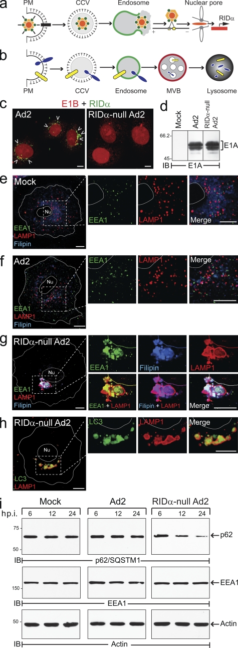
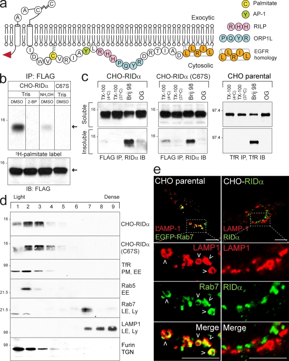
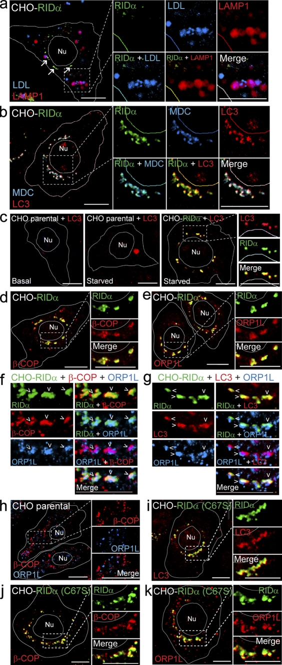
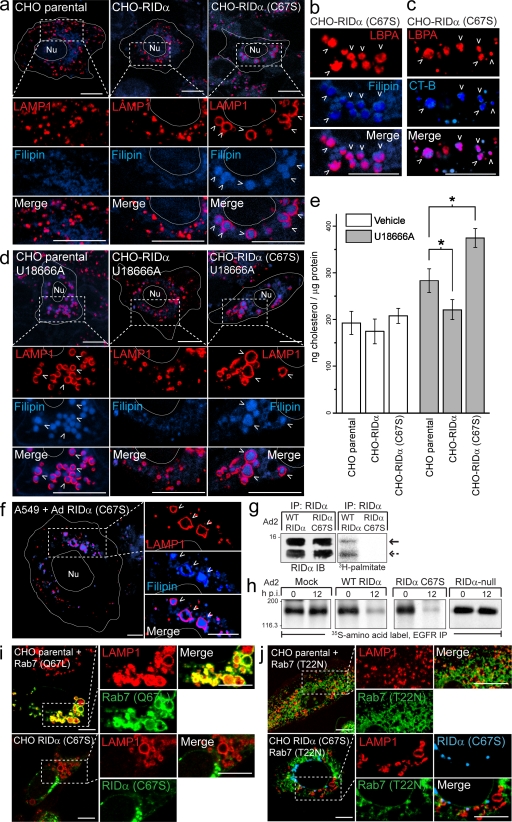
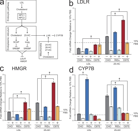
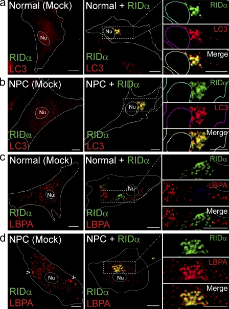
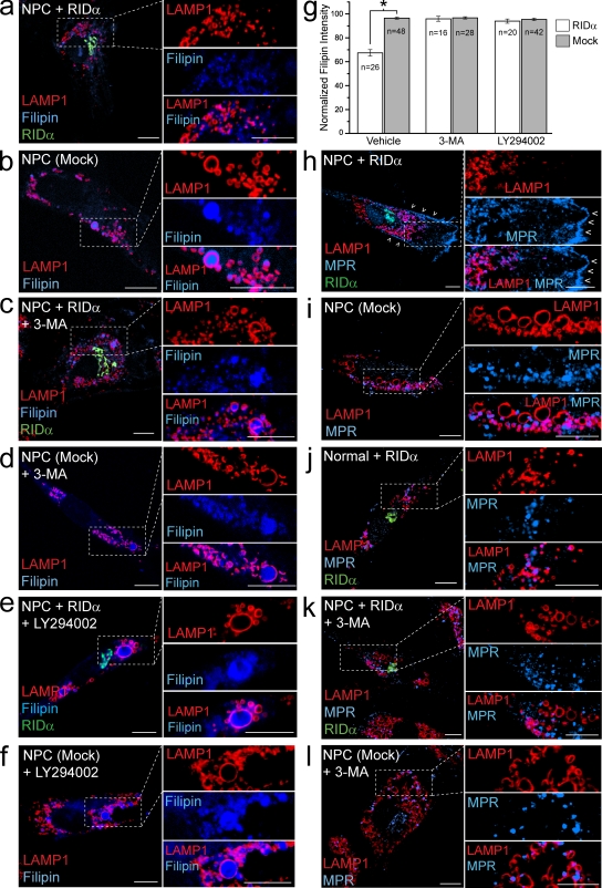
Host-pathogen interactions are important model systems for understanding fundamental cell biological processes. In this study, we describe a cholesterol-trafficking pathway induced by the adenovirus membrane protein RID-alpha that also subverts the cellular autophagy pathway during early stages of an acute infection. A palmitoylation-defective RID-alpha mutant deregulates cholesterol homeostasis and elicits lysosomal storage abnormalities similar to mutations associated with Niemann-Pick type C (NPC) disease. Wild-type RID-alpha rescues lipid-sorting defects in cells from patients with this disease by a mechanism involving a class III phosphatidylinositol-3-kinase. In contrast to NPC disease gene products that are localized to late endosomes/lysosomes, RID-alpha induces the accumulation of autophagy-like vesicles with a unique molecular composition. Ectopic RID-alpha regulates intracellular cholesterol trafficking at two distinct levels: the egress from endosomes and transport to the endoplasmic reticulum necessary for homeostatic gene regulation. However, RID-alpha also induces a novel cellular phenotype, suggesting that it activates an autonomous cholesterol regulatory mechanism distinct from NPC disease gene products.


Figures

Figure 1.
Figure 1.
RID-α counterbalances Ad-induced autophagy. (a) Ad2 internalized by clathrin-mediated endocytosis escapes from EEs by membrane lysis followed by MT-dependent transport to the nucleus and viral gene expression. (b) RID-α, which is expressed within the first few hours of an acute infection, facilitates targeted degradation of EGFRs (blue) and proapoptotic receptors (yellow). (c) Confocal images of A549 cells infected with wild-type or RID-α–null Ad2 viruses stained with E1B and RID-α antibodies. Arrowheads indicate RID-α–positive compartments. (d) Equal aliquots of total cellular protein from mock- and Ad-infected A549 cells resolved by SDS-PAGE and immunoblotted with E1A antibody. (e–g) Confocal images of mock (e), Ad2 (f), and RID-α–null Ad2-infected (g) A549 cells stained with EEA1 and LAMP1 antibodies and filipin; magnified images of single and merged channels are shown on the right. (h) Confocal images of RID-α–null Ad2-infected A549 cells stained for LC3 and LAMP1. (e–h) Cell and nucleus (Nu) boundaries were drawn using MetaMorph software. Boxed areas show regions of the image that were magnified. (i) Equal aliquots of total cellular protein from mock- and Ad-infected cells immunoblotted with antibodies to p62/SQSTM1, EEA1, and actin as a function of time postinfection (p.i.). CCV, clathrin-coated vesicle; IB, immunoblot. (d and i) Molecular mass is indicated in kilodaltons. Bars, 10 µm.
Figure 2.
Figure 2.
RID-α is palmitoylated at Cys67. (a) Schematic of RID-α membrane topology showing the amino-terminal Flag epitope (red arrowhead), luminal signal peptidase cleavage (which generates 13.7- or 11.3-kD RID-α species) and disulfide bond formation sites, carboxyl tail sequence with palmitoylation and protein interaction sites, and the EGFR homology domain (Hoffman et al., 1992b; Vinogradova et al., 1998; Tsacoumangos et al., 2005; Cianciola et al., 2007; Shah et al., 2007). (b) CHO–RID-α and CHO–RID-α (C67S) cells treated with 2-BP or DMSO and radiolabeled with [3H]palmitate. Flag immunocomplexes were separated by SDS-PAGE, and gels were incubated with Tris or hydroxylamine solutions before fluorography. The bottom panel shows Flag immunocomplexes from duplicate samples analyzed by RID-α immunoblotting for loading control. Arrows denote 13.7-kD and RID-α species. (c) CHO cell lines extracted with the indicated detergents and RID-α (left and middle) or TfR (right) immunocomplexes from detergent-soluble and -insoluble fractions immunoblotted with antibodies to the same protein. (d) Membranes from stable CHO cells expressing wild-type RID-α or RID-α (C67S) fractionated on 27% Percoll gradients and equal aliquots of total membrane protein immunoblotted with antibodies to RID-α (top two panels) are shown. Equal aliquots of total membrane protein from parental CHO cells immunoblotted with antibodies to marker proteins for intracellular membrane compartments listed in the figure (bottom five panels) are shown. (b–d) Molecular mass is indicated in kilodaltons. (e) Confocal images of CHO cells transfected with EGFP-Rab7 plasmid and stained for LAMP1 or CHO–RID-α cells stained for LAMP1 and RID-α. Boxed areas show regions of the image that were magnified. Arrowheads denote compartments with colocalized markers. IB, immunoblot; IP, immunoprecipitation; Ly, lysosome; OG, octyl glucoside; TX-100, Triton X-100. Bars, 10 µm.
Figure 3.
Figure 3.
RID-α induces formation of a hybrid organelle with characteristics of both endocytic and autophagic vesicles. (a) CHO–RID-α cells stained with LAMP1 and RID-α antibodies after incubation with DiI-LDL. Arrows denote the RID-α compartment. (b) CHO–RID-α cells stained for LC3 and RID-α after incubation with MDC. (c) Parental CHO or CHO–RID-α cells stained for LC3 or LC3 and RID-α under basal conditions (left) or after 6-h incubation in Earl’s balanced salt solution to induce autophagy (middle and right). (d and e) CHO–RID-α cells stained for RID-α and β-COP (d) or ORP1L (e). (f and g) Magnified images of single and merged channels of CHO–RID-α cells triple stained for RID-α and ORP1L and either β-COP (f) or LC3 (g). Arrowheads denote compartments with colocalized markers. (h) Parental CHO cells stained for β-COP and ORP1L. (i–k) CHO–RID-α (C67S) cells stained for RID-α and LC3 (i), β-COP (j), or ORP1L (k). (a–e and h–k) Cell and nucleus (Nu) boundaries were drawn using MetaMorph software. Boxed areas show regions of the image that were magnified. Bars, 10 µm.
Figure 4.
Figure 4.
RID-α (C67S) induces the formation of enlarged lipid-filled LAMP1 structures. (a) Confocal images of CHO cell lines stained with LAMP1 antibody and filipin. (b and c) Magnified images of single and merged channels from CHO–RID-α (C67S) cells stained with LBPA antibody and filipin (b) or LBPA antibody after incubation with Alexa Fluor 647 CT-B (c). (d) CHO cell lines treated with U18666A for 8 h and stained with LAMP1 antibody and filipin. (e) Cholesterol quantification in CHO cell lines treated with DMSO (vehicle) or U18666A for 8 h using the Amplex red cholesterol assay kit. Values were normalized to total cellular protein and are displayed as mean ± SEM (*, P < 0.01). (f) Confocal images of A549 cells infected with a mutant RID-α (C67S) Ad2 virus and stained with LAMP1 antibody and filipin 24 h postinfection. (g) A549 cells infected with wild-type (WT) or RID-α (C67S) Ad2 viruses radiolabeled with [3H]palmitate and RID-α immunocomplexes separated by SDS-PAGE for fluorography. Arrows denote 13.7 (solid)- or 11.3 (dashed)-kD RID-α species. (h) A549 cells radiolabeled with 35S-Express Protein Labeling mix and mock infected or infected with wild-type or mutant Ad viruses and EGFR immunocomplexes analyzed by SDS-PAGE and fluorography at the times indicated. (g and h) Molecular mass is indicated in kilodaltons. (i) Parental CHO cells transfected with constitutively active (Q67L) EGFP-Rab7 and stained for LAMP1 or CHO–RID-α (C67S) cells stained for LAMP1 and RID-α. (j) Parental CHO or CHO–RID-α (C67S) cells transfected with dominant-negative (T22N) EGFP-Rab7 and stained for LAMP1 (parental) or LAMP1 and RID-α (CHO–RID-α (C67S)). (a–d and f) Arrowheads indicate examples of costained vesicles. (a, d, f, i, and j) Boxed areas show regions of the image that were magnified. (a, d, and f) Cell and nucleus (Nu) boundaries were drawn using MetaMorph software. IB, immunoblot; IP, immunoprecipitation; p.i., postinfection. Bars, 10 µm.
Figure 5.
Figure 5.
RID-α modulates sterol-regulated gene expression. (a) Transcriptional mechanisms controlling expression of the target genes LDLR, HMGR, and CYP7B described in Results. (b–d) LDLR (b), HMGR (c), and CYP7B (d) mRNA levels quantified by real-time PCR. Values are expressed as relative units after internal normalization to glyceraldehyde 3-phosphate dehydrogenase mRNA levels and compared with control samples from the same cell lines cultured in 10% FBS from three independent experiments. Data are presented as mean ± SEM (*, P < 0.001).
Figure 6.
Figure 6.
RID-α induces formation of dynamic hybrid organelles with characteristics of both endocytic and autophagic vesicles in NPC fibroblasts. (a and b) Confocal images of normal (a) or NPC cells (b) stained for RID-α and LC3 after mock transfection or transfection with a RID-α expression plasmid. (c and d) Confocal images of normal (c) or NPC cells (d) stained for RID-α and LBPA after mock transfection or transfection with a RID-α expression plasmid. (d) Arrowheads show presumptive LBPA-positive LSOs. (a–d) Cell and nucleus (Nu) boundaries were drawn using MetaMorph software. Boxed areas show regions of the image that were magnified. Bars, 10 µm.
Figure 7.
Figure 7.
RID-α expression rescues cholesterol trafficking and associated defects in NPC cells by a class III PI3K–dependent mechanism. (a–f) NPC cells transfected in the absence or presence of PI3K inhibitors, as indicated, and stained for LAMP1, filipin, and RID-α (a, c, and e) or LAMP1 and filipin (b, d, and f). (g) Quantification of normalized filipin fluorescence intensity in cells treated similarly as in a–f and as described in Materials and methods. Data are presented as mean ± SEM (*, P < 0.001). (h–l) NPC and normal cells transfected in the absence or presence of PI3K inhibitors, as indicated, and stained for LAMP1, MPR, and RID-α (h, j, and k) or LAMP1 and MPR (i and l). Cells in h–l were cultured in 10% LPDS media for 3 d followed by 50 µg/ml LDL for 24 h before staining. (h) Arrowheads denote PM localization of MPR. (a–f and h–l) Boxed areas show regions of the image that were magnified. Bars, 10 µm.
Figure 8.
Figure 8.
Model of the role of RID-α as a coordinator of endosome trafficking. LDL-cholesterol (Chol) esters are internalized via the PM LDLR (1a), unesterified, and trafficked to MVBs/LEs (1b). Cholesterol is egressed out of these organelles by the coordinated action of NPC1/NPC2 and transported to the ER, where it is sensed by cholesterol and 25-HC homeostatic machinery (2a), and to mitochondria, where it is converted to other oxysterols (2b). NPC1/NPC2 mutations block cholesterol egress to both of these compartments, deregulate cholesterol homeostasis, reduce oxysterol production, and induce LSO formation (3). Although RID-α vesicles resemble autophagic vesicles, they also have distinct molecular properties that distinguish them from bona fide autophagosomes (4a). Specialized RID-α compartments that sequester the MDC indicator dye from bulk cytosol may supply an unknown rate-limiting factor important for endocytic maintenance and/or cholesterol homeostasis. RID-α activates an autonomous class III PI3K–dependent cholesterol egress mechanism that restores ER cholesterol trafficking in NPC1-defective cells (4b). RID-α also suppresses Ad-induced autophagy (5a) and facilitates endosome to lysosome targeting of select membrane protein cargo (5b). Positive and negative RID-α–mediated actions are highlighted in red.

Similar articles

Cited by

References

    1. Backer J.M. 2008. The regulation and function of Class III PI3Ks: novel roles for Vps34. Biochem. J. 410:1–17 10.1042/BJ20071427 - DOI - PubMed
    1. Biederbick A., Kern H.F., Elsässer H.P. 1995. Monodansylcadaverine (MDC) is a specific in vivo marker for autophagic vacuoles. Eur. J. Cell Biol. 66:3–14 - PubMed
    1. Bright N.A., Gratian M.J., Luzio J.P. 2005. Endocytic delivery to lysosomes mediated by concurrent fusion and kissing events in living cells. Curr. Biol. 15:360–365 10.1016/j.cub.2005.01.049 - DOI - PubMed
    1. Brown D.A., Rose J.K. 1992. Sorting of GPI-anchored proteins to glycolipid-enriched membrane subdomains during transport to the apical cell surface. Cell. 68:533–544 10.1016/0092-8674(92)90189-J - DOI - PubMed
    1. Bucci C., Thomsen P., Nicoziani P., McCarthy J., van Deurs B. 2000. Rab7: a key to lysosome biogenesis. Mol. Biol. Cell. 11:467–480 - PMC - PubMed

Publication types

MeSH terms