. 2010 Jan 29;285(5):2976-85.
doi: 10.1074/jbc.M109.038398. Epub 2009 Nov 30.

The linker for activation of B cells (LAB)/non-T cell activation linker (NTAL) regulates triggering receptor expressed on myeloid cells (TREM)-2 signaling and macrophage inflammatory responses independently of the linker for activation of T cells

Affiliations

The linker for activation of B cells (LAB)/non-T cell activation linker (NTAL) regulates triggering receptor expressed on myeloid cells (TREM)-2 signaling and macrophage inflammatory responses independently of the linker for activation of T cells

Gillian C Whittaker et al. J Biol Chem. .

Abstract

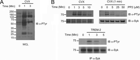
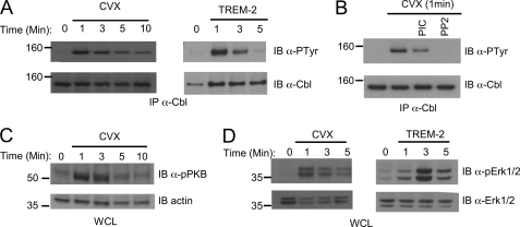
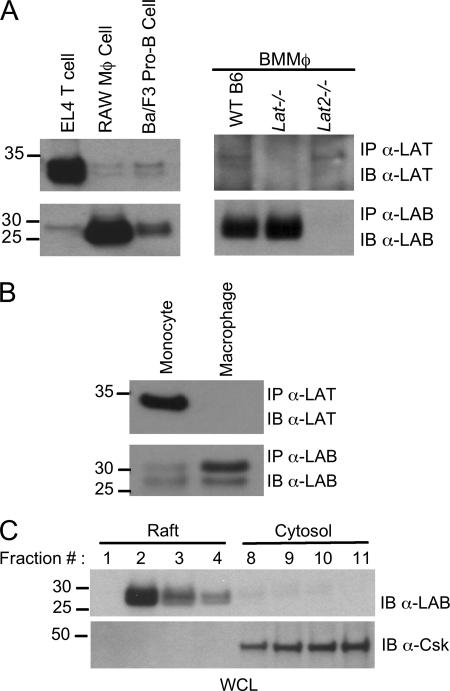
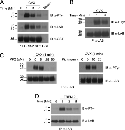
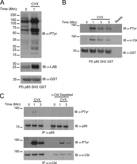
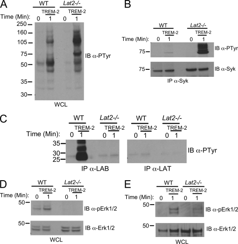
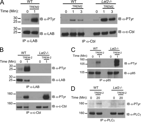
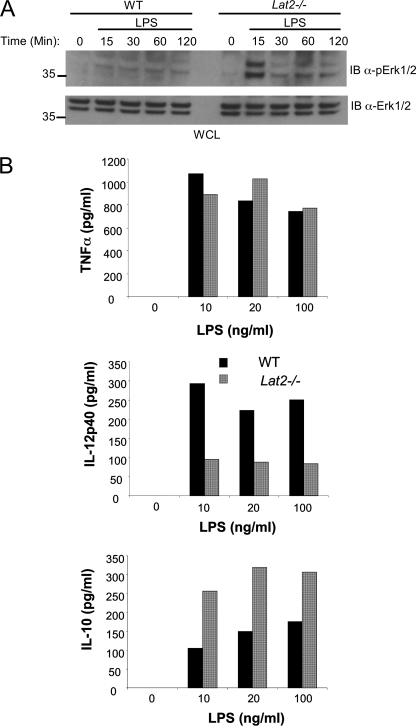
Triggering receptor expressed on myeloid cells-2 (TREM-2) is rapidly emerging as a key regulator of the innate immune response via its regulation of macrophage inflammatory responses. Here we demonstrate that proximal TREM-2 signaling parallels other DAP12-based receptor systems in its use of Syk and Src-family kinases. However, we find that the linker for activation of T cells (LAT) is severely reduced as monocytes differentiate into macrophages and that TREM-2 exclusively uses the linker for activation of B cells (LAB encoded by the gene Lat2(-/-)) to mediate downstream signaling. LAB is required for TREM-2-mediated activation of Erk1/2 and dampens proximal TREM-2 signals through a novel LAT-independent mechanism resulting in macrophages with proinflammatory properties. Thus, Lat2(-/-) macrophages have increased TREM-2-induced proximal phosphorylation, and lipopolysaccharide stimulation of these cells leads to increased interleukin-10 (IL-10) and decreased IL-12p40 production relative to wild type cells. Together these data identify LAB as a critical, LAT-independent regulator of TREM-2 signaling and macrophage development capable of controlling subsequent inflammatory responses.


Figures

FIGURE 1.
FIGURE 1.
Proximal TREM-2 signaling. A, whole cell lysates (WCL) from Cvx-stimulated chimera-expressing RAW cells were immunoblotted (IB) with an anti-phosphotyrosine (clone 4G10) antibody. B, top, chimera expressing RAW264.7 cells were treated for 30 min with various concentrations of the Src inhibitor PP2 as indicated (right panel) or left untreated (left panel). Cells were stimulated with Cvx for the indicated times, and Syk was immunoprecipitated, resolved via SDS-PAGE, and immunoblotted with anti-phosphotyrosine. Filters were reprobed with anti-Syk as indicated to demonstrate equal loading. B, bottom. BMMΦ of WT mice were stimulated for the times indicated with TREM-2 monoclonal antibody, and Syk phosphorylation was assayed as above.
FIGURE 2.
FIGURE 2.
Distal TREM-2 signaling events. A, chimera expressing RAW264.7 cells (left) or BMMΦ from WT mice (right) were stimulated with 20 nm Cvx or anti-TREM-2, respectively, for the indicated times. Lysates were immunoprecipitated (IP) with anti-c-Cbl and immunoblotted with anti-phosphotyrosine. Blots were stripped and reprobed with an anti-c-Cbl. B, before stimulation with Cvx, chimera-expressing cells were treated for 30 min with 20 μg/ml piceatannol or 25 μm PP2. Lysates were immunoprecipitated with an anti-c-Cbl and immunoblotted with an anti-phosphotyrosine. Blots were stripped and reprobed with anti-c-Cbl. C, chimera expressing RAW264.7 cells were stimulated with 20 nm Cvx as in A. Lysates were immunoblotted with an anti-phosphoprotein kinase B. Reprobing with anti-actin demonstrated equal loading. WCL, whole cell lysates. D, chimera-expressing cells (left) or BMMΦ from WT mice (right) were stimulated as in A, and lysates were immunoblotted with anti-phospho-Erk1/2. Blots were stripped and reprobed with anti-Erk1/2, as indicated. Results are representative of at least two independent experiments.
FIGURE 3.
FIGURE 3.
LAT and LAB expression in monocytes and macrophages. Equal amounts of protein derived from lysates of EL4 T cells, RAW264.7, Ba/F3 B cells, WT, Lat−/−, and Lat2−/− BMMΦ (A) or human monocytes or macrophages differentiated with M-CSF (B) were immunoprecipitated with anti-LAT or anti-LAB antibodies as indicated. The corresponding immunoprecipitates (IP) were subject to immunoblotting (IB) with anti-LAT or anti-LAB monoclonal antibodies. C, detergent-insoluble raft fractions were isolated by sucrose-density gradient ultracentrifugation from RAW264.7 lysates. Raft fractions (1–4) and non-raft fractions (9–11) were immunoblotted with an anti-LAB antibody or anti-Csk antibody. Results are representative of at least two independent experiments. WCL, whole cell lysates.
FIGURE 4.
FIGURE 4.
LAB is tyrosine-phosphorylated downstream of TREM-2. A–C, chimera expressing RAW264. 7 cells were stimulated for the indicated times with 20 nm Cvx. A, pulldown assays were performed on postnuclear lysates using Grb-2 SH2 domain fusion protein then immunoblotted (IB) with an anti-phosphotyrosine. The same blot was stripped and reprobed with anti-LAB antibody followed by anti-GST. B, Cvx-stimulated lysates were immunoprecipitated (IP) with anti-LAB antibody and immunoblotted with an anti-phosphotyrosine. The same blot was stripped and reprobed with an anti-LAB. C, before stimulation with Cvx, samples were treated for 30 min with various concentrations of the PP2 or piceatannol, as indicated. D, BMMΦ from WT mice were stimulated for the times indicated by TREM-2 cross-linking. Lysates were immunoprecipitated with anti-LAB antibody and immunoblotted with an anti-phosphotyrosine. The same blots were stripped and reprobed with an anti-LAB antibody. Results are representative of several independent experiments.
FIGURE 5.
FIGURE 5.
LAB is present in a complex with p85 and c-Cbl after TREM-2 stimulation. A, GST pulldown (PD) assays from Cvx-stimulated RAW264.7 cell lysates were performed using a p85 SH2 domain GST fusion protein. Protein complexes were resolved by SDS-PAGE and immunoblotted (IB) with anti-phosphotyrosine (top), anti-LAB antibody (middle), or anti-GST (bottom). B, after Cvx stimulation for the indicated times lysates were precipitated with empty beads (lane 5) or GST-p85 SH2 (lanes 1–4). Complexes were resolved as above and immunoblotted with anti-phosphotyrosine (top), anti-c-Cbl (middle), or anti-GST (bottom). C, Cvx-stimulated lysates were subjected to three rounds of immunodepletion with anti-c-Cbl. Cbl-depleted lysates were subsequently immunoprecipitated with either anti-p85 (top two panels) or anti-c-Cbl (bottom two panels). Immunoblots were probed with anti-phosphotyrosine and then stripped and reprobed with anti-p85 or anti-c-Cbl as appropriate. Results are representative of at least two independent experiments.
FIGURE 6.
FIGURE 6.
LAB regulates TREM-2 signaling in macrophages independently of LAT. A, immortalized MΦ from WT or Lat2−/− mice were stimulated by TREM-2 cross-linking for the times indicated. Equal amounts of protein were immunoblotted (IB) with anti-phosphotyrosine. WCL, whole cell lysates. B, lysates as in A were immunoprecipitated (IP) with anti-Syk and immunoblotted with an anti-phosphotyrosine. The same blot was stripped and reprobed with an anti-Syk. Results are representative of at least three independent experiments. C, equal amounts of protein from TREM-2-stimulated lysates of BMMΦ from WT or Lat2−/− mice were immunoprecipitated with anti-LAB or anti-LAT antibodies. Blots were probed with anti-phosphotyrosine, and both panels shown here are from the same exposure. D, immortalized MΦ from WT and Lat2−/− mice were stimulated with TREM-2 and immunoblotted with an anti-phospho Erk1/2 antibody. The same blot was stripped and reprobed with anti- Erk1/2. Results are representative of at least two independent experiments. E, BMMΦ derived from WT or Lat2−/− mice were assayed for Erk1/2 activation as in D.
FIGURE 7.
FIGURE 7.
Enhanced c-Cbl phosphorylation in the absence of LAB. A, immortalized MΦ from WT and Lat2−/− mice were stimulated by TREM-2 cross-linking for the indicated times. Lysates were immunoprecipitated (IP) with an anti-LAB polyclonal antibody (left) or an anti-c-Cbl antibody (right). Immunoblots (IB) were probed with anti-phosphotyrosine, anti-LAB, or anti-c-Cbl as indicated. B, BMMΦ from the indicated strain were stimulated, immunoprecipitated, and immunoblotted as in A. C, lysates from TREM-2-stimulated immortalized MΦ were immunoprecipitated with an anti-p85 and immunoblotted with an anti-phosphotyrosine. The same blot was stripped and reprobed with an anti-p85. D, lysates from TREM-2-stimulated immortalized MΦ were immunoprecipitated with an anti-PLCγ and immunoblotted with an anti-phosphotyrosine. The same blot was stripped and reprobed with an anti-PLCγ. Results are representative of several independent experiments.
FIGURE 8.
FIGURE 8.
LAB differentially regulates production of IL-10 and IL-12p40 by macrophages. A, BMMΦ from WT and Lat2−/− mice were stimulated for the indicated times with LPS, and phospho-Erk was assessed by immunoblotting (IB) whole cell lysates. B, BMMΦ from WT and Lat2−/− mice were stimulated for 24 h with various concentrations of LPS as indicated. Supernatants were quantified for IL-10, IL-12p40, and TNFα by enzyme-linked immunosorbent assay. Black bars represent WT, and gray bars represent Lat2−/−. Results are representative of at least three independent experiments with a pool of three mice in each.

Similar articles

Cited by

References

    1. Ford J. W., McVicar D. W. (2009) Curr. Opin. Immunol. 21, 38–46 - PMC - PubMed
    1. Bouchon A., Dietrich J., Colonna M. (2000) J. Immunol. 164, 4991–4995 - PubMed
    1. Bouchon A., Facchetti F., Weigand M. A., Colonna M. (2001) Nature 410, 1103–1107 - PubMed
    1. Schenk M., Bouchon A., Seibold F., Mueller C. (2007) J. Clin. Invest. 117, 3097–3106 - PMC - PubMed
    1. Bouchon A., Hernández-Munain C., Cella M., Colonna M. (2001) J. Exp. Med. 194, 1111–1122 - PMC - PubMed

Publication types

MeSH terms