. 2010 Jan 22;285(4):2318-25.
doi: 10.1074/jbc.M109.071498. Epub 2009 Nov 30.

Inducible priming phosphorylation promotes ligand-independent degradation of the IFNAR1 chain of type I interferon receptor

Affiliations

Inducible priming phosphorylation promotes ligand-independent degradation of the IFNAR1 chain of type I interferon receptor

Sabyasachi Bhattacharya et al. J Biol Chem. .

Abstract

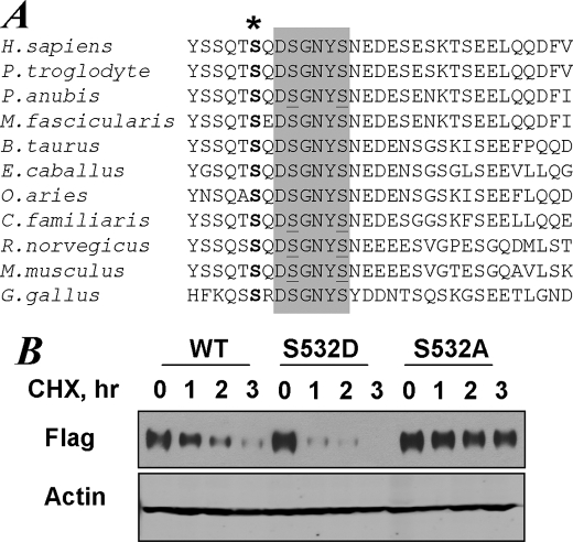
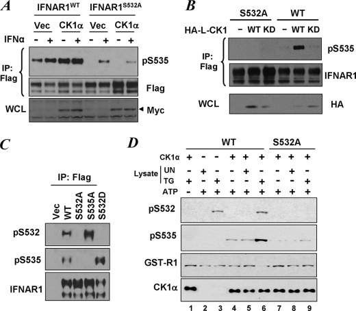
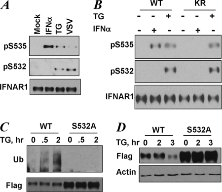
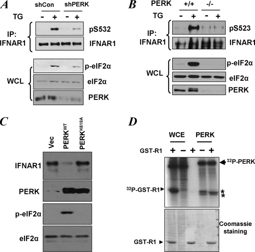
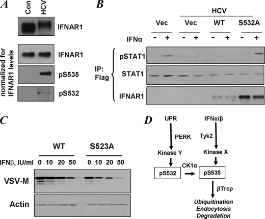
Phosphorylation-dependent ubiquitination and ensuing down-regulation and lysosomal degradation of the interferon alpha/beta receptor chain 1 (IFNAR1) of the receptor for Type I interferons play important roles in limiting the cellular responses to these cytokines. These events could be stimulated either by the ligands (in a Janus kinase-dependent manner) or by unfolded protein response (UPR) inducers including viral infection (in a manner dependent on the activity of pancreatic endoplasmic reticulum kinase). Both ligand-dependent and -independent pathways converge on phosphorylation of Ser(535) within the IFNAR1 degron leading to recruitment of beta-Trcp E3 ubiquitin ligase and concomitant ubiquitination and degradation. Casein kinase 1 alpha (CK1 alpha) was shown to directly phosphorylate Ser(535) within the ligand-independent pathway. Yet given the constitutive activity of CK1 alpha, it remained unclear how this pathway is stimulated by UPR. Here we report that induction of UPR promotes the phosphorylation of a proximal residue, Ser(532), in a pancreatic endoplasmic reticulum kinase-dependent manner. This serine serves as a priming site that promotes subsequent phosphorylation of IFNAR1 within its degron by CK1 alpha. These events play an important role in regulating ubiquitination and degradation of IFNAR1 as well as the extent of Type I interferon signaling.


Figures

FIGURE 1.
FIGURE 1.
The conserved priming site within IFNAR1 regulates the intrinsic stability of the protein. A, alignment of primary sequences of IFNAR1 from indicated species including Homo sapiens, Pan troglodyte, Papia anubis, Macaca fascicularis, Bos taurus, Equus caballus, Ovis aries, Canis familiaris, Rattus norvegicus, Mus musculus, and Gallus gallus. The phospho-degron sequences are shaded, and serine residues within the degron are underlined. The conserved putative priming site (Ser532 in human IFNAR1) is denoted by an asterisk. B, degradation of FLAG-IFNAR1 (wild type (WT) or S532A mutant) overexpressed in 293T cells was analyzed by cycloheximide (CHX, 2 mm) chase for the indicated times followed by immunoblotting using anti-FLAG antibody. The levels of β-actin were also analyzed as a loading control.
FIGURE 2.
FIGURE 2.
Priming phosphorylation is required for the ligand-independent phosphorylation of IFNAR1 degron. A, degron phosphorylation of FLAG-IFNAR1 (wild type (WT) or S532A mutant) co-expressed in 293T cells with Myc-tagged human CK1α or empty vector (Vec) and treated or not with IFNα (1000 IU/ml for 30 min as indicated) was analyzed by FLAG immunoprecipitation (IP) followed by immunoblotting using the indicated antibodies. The levels of Myc-CK1α in whole cell lysates were also determined. B, FLAG-IFNAR1 (wild type or S532A mutant) was co-expressed in 293T cells with HA-tagged Leishmania CK1 (HA-L-CK1, wild type, or kinase dead (KD)) and purified by FLAG immunoprecipitation. Phosphorylation of the IFNAR1 degron and levels of IFNAR1 were analyzed by immunoblotting using the indicated antibodies. The levels of HA-L-CK1 in whole cell lysates (WCL) were also determined. C, characterization of anti-Ser(P)532 antibody. FLAG-IFNAR1 proteins (wild type, S535A, or S532A mutants) were expressed in 293T cells, immunopurified, and analyzed using the indicated antibodies. Vec, reactions from cells transfected with empty vector (pCDNA3). D, 293T cells were left untreated (UN) or were treated with TG (1 μm for 30 min) and harvested. Lysates from these cells were twice immunodepleted with antibodies against CK1α, and the CK1α-free supernatants (4 μg) were used alone (lanes 2 and 3) or together with 0.5 μg of bacterially produced recombinant GST-CK1α (lanes 1 and 4–9) for in vitro phosphorylation of GST-IFNAR1 (wild type, lanes 1–6, or S532A mutant, lanes 7–9) in the presence of ATP (except in lane 1) at 30 °C for 30 min as indicated. Phosphorylation of GST-IFNAR1 on Ser532 and Ser535, levels of GST-IFNAR1 (using anti-GST antibody), and levels of CK1α were analyzed by immunoblotting.
FIGURE 3.
FIGURE 3.
UPR induces phosphorylation of the priming site that is required for increased ubiquitination and degradation of IFNAR1. A, endogenous IFNAR1 proteins immunopurified from HeLa cells that were untreated (Mock) or were treated with TG (1 μm for 30 min), IFNα (6000 IU/ml for 30 min), or infected with VSV (0.1 multiplicity of infection for 1 h followed by an additional 10 h of incubation in virus-free medium) as indicated, were analyzed by immunoblotting using the indicated antibodies. B, endogenous IFNAR1 proteins immunopurified from 11.1-Tyk2-null derivative cell lines reconstituted with wild type Tyk2 (WT) or catalytically deficient Tyk2 (KR) were treated with TG or IFNα (in doses indicated in A) for 30 min. C, FLAG-IFNAR1 proteins (wild type or S532A mutant) were expressed in 293T cells. The cells were pretreated with a lysosomal inhibitor (methylamine HCl, 10 mm) for 1 h to prevent degradation of ubiquitinated receptors. Then the cells were treated with TG (1 μm for the indicated times), and FLAG-IFNAR1 proteins were immunopurified under denaturing conditions and analyzed by immunoblotting using antibodies against ubiquitin (Ub, upper panel) and FLAG (lower panel). D, down-regulation of the levels of FLAG-IFNAR1 (wild type or S532A mutant) expressed in 293T cells treated with TG (1 μm for indicated times) was analyzed by immunoblotting using FLAG antibody. Comparable loading was verified by anti-β-actin immunoblot (lower panel).
FIGURE 4.
FIGURE 4.
Role of PERK in UPR-induced phosphorylation of priming site of IFNAR1. A, HeLa cells transfected with small hairpin RNA against PERK or against GFP (shCon) were treated with TG (1 μm for 30 min), and endogenous IFNAR1 proteins were immunopurified (IP) and analyzed for phosphorylation on the priming site and for total levels by immunoblotting using the indicated antibodies. Phosphorylation of a known PERK substrate eIF2α (as well as its total levels) and the levels of PERK itself were also determined in whole cell lysates (WCL). B, mouse embryo fibroblasts from wild type or PERK knock-out animals were treated with TG as indicated. The levels and priming phosphorylation of endogenous murine IFNAR1 on Ser523 (analogue of human Ser532) were analyzed by immunoblotting using indicated antibodies. The phosphorylation and levels of eIF2α and the levels of PERK in whole cell lysates were also determined. C, levels of endogenous IFNAR1 in 293T cells transfected with wild type or the catalytically deficient mutant (K618A) of PERK were analyzed by immunoprecipitation followed by immunoblotting using an anti-IFNAR1 antibody. The levels of PERK, phosphorylated PERK, and levels of eIF2α were also examined. D, whole cell extracts (WCE) from 293T cells or recombinant bacterially produced constitutively active ΔN-PERK were incubated alone or with GST-IFNAR1 in the presence of radiolabeled [γ-32P]ATP as indicated. Resulting phosphorylation of GST-IFNAR1 or contaminants and autophosphorylation of PERK was determined by SDS-PAGE followed by Coomassie staining and autoradiography. The positions of PERK, GST-IFNAR1, and some irrelevant contaminants (denoted by asterisks) are indicated. Vec, empty vector.
FIGURE 5.
FIGURE 5.
Priming phosphorylation of IFNAR1 contributes to regulation of the extent of IFNα/β signaling. A, control (Con) human Huh7 cells and those expressing the HCV replicon were analyzed for IFNAR1 levels by immunoprecipitation-immunoblotting (upper panel). The lower three panels depict the experiments where gel loading was normalized to achieve comparable levels of immunopurified IFNAR1 in each lane. Phosphorylation of IFNAR1 on Ser532 and Ser535 was determined by immunoblotting using the indicated antibodies. B, control human Huh7 cells and those expressing the HCV replicon were transfected with FLAG-tagged STAT1 alone with empty vector (Vec) or FLAG-IFNAR1 (wild type (WT) or S532A mutant) and were untreated or treated with IFNα (60 IU/ml for 30 min) as indicated. Lysates of these cells were immunoprecipitated (IP) using anti-FLAG antibody and analyzed by immunoblotting using antibodies against phospho-STAT1, total STAT1, and IFNAR1. C, mouse embryo fibroblasts from IFNAR1-null animals were reconstituted with murine FLAG-IFNAR1 (wild type or S523A mutant, which is a mouse analogue of human S532A mutant). The cells were treated with indicated doses of murine IFNβ for 1 h, incubated for 8 h in fresh medium, and then infected with VSV (multiplicity of infection of 0.1). Expression of VSV-M protein was analyzed 16 h later by immunoblotting. Levels of β-actin were also determined (lower panel). D, model for ligand-dependent and ligand-independent ubiquitination and degradation of IFNAR1. Both pathways converge at the level of degron phosphorylation (Ser(P)535, pS535). Signaling induced by IFN and dependent on the activity of Tyk2 does not require either CK1α (24) or priming phosphorylation (this study). Ligand-independent pathway initiated by inducers of UPR does not need either ligand or Tyk2 activity but requires CK1α (26) and PERK-dependent priming phosphorylation (this study).

References

    1. Wallach D. F. (1987) Fundamentals of Receptor Molecular Biology, M. Dekker, New York
    1. Müller U., Steinhoff U., Reis L. F., Hemmi S., Pavlovic J., Zinkernagel R. M., Aguet M. (1994) Science. 264, 1918–1921 - PubMed
    1. Gutterman J. U. (1994) Proc. Natl. Acad. Sci. U.S.A. 91, 1198–1205 - PMC - PubMed
    1. Billard C., Sigaux F., Castaigne S., Valensi F., Flandrin G., Degos L., Falcoff E., Aguet M. (1986) Blood. 67, 821–826 - PubMed
    1. Maxwell B. L., Talpaz M., Gutterman J. U. (1985) Int. J. Cancer 36, 23–28 - PubMed

Publication types

MeSH terms