. 2010 Jan 29;285(5):2986-95.
doi: 10.1074/jbc.M109.070466. Epub 2009 Nov 30.

A new isoquinolinium derivative, Cadein1, preferentially induces apoptosis in p53-defective cancer cells with functional mismatch repair via a p38-dependent pathway

Affiliations

A new isoquinolinium derivative, Cadein1, preferentially induces apoptosis in p53-defective cancer cells with functional mismatch repair via a p38-dependent pathway

Eun Ryoung Jang et al. J Biol Chem. .

Abstract

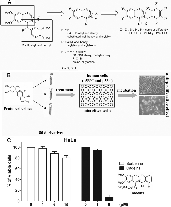
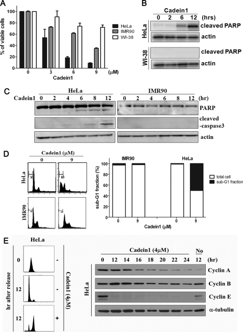
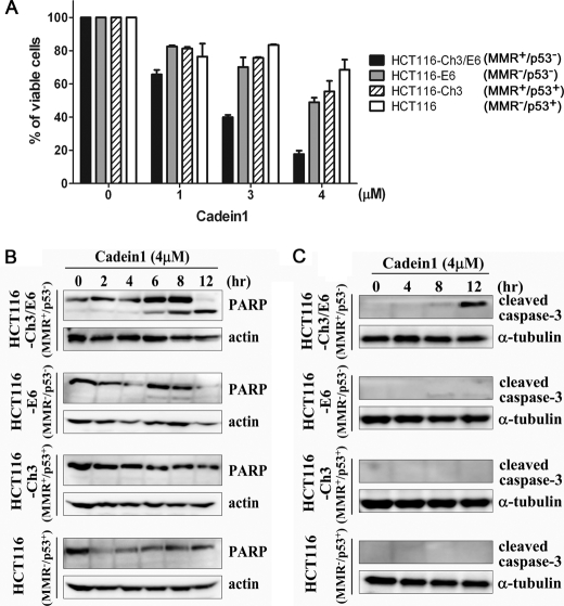
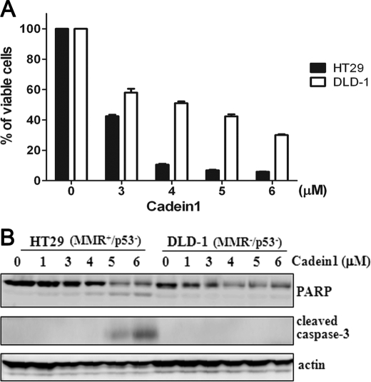
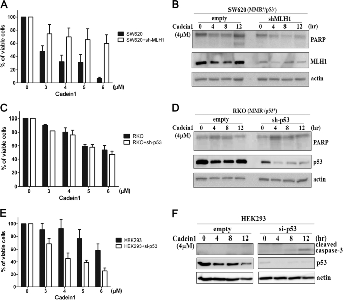
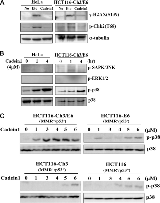
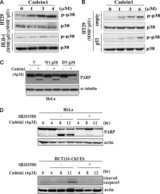
We screened a protoberberine backbone derivative library for compounds with anti-proliferative effects on p53-defective cancer cells. A compound identified from this small molecule library, cadein1 (cancer-selective death inducer 1), an isoquinolinium derivative, effectively leads to a G(2)/M delay and caspase-dependent apoptosis in various carcinoma cells with non- functional p53. The ability of cadein1 to induce apoptosis in p53-defective colon cancer cells was tightly linked to the presence of a functional DNA mismatch repair (MMR) system, which is an important determinant in chemosensitivity. Cadein1 was very effective in MMR(+)/p53(-) cells, whereas it was not effective in p53(+) cells regardless of the MMR status. Consistently, when the function of MMR was blocked with short hairpin RNA in SW620 (MMR(+)/p53(-)) cells, cadein1 was no longer effective in inducing apoptosis. Besides, the inhibition of p53 increased the pro-apoptotic effect of cadein1 in HEK293 (MMR(+)/p53(+)) cells, whereas it did not affect the response to cadein1 in RKO (MMR(-)/p53(+)) cells. The apoptotic effects of cadein1 depended on the activation of p38 but not on the activation of Chk2 or other stress-activated kinases in p53-defective cells. Taken together, our results show that cadein1 may have a potential to be an anti-cancer chemotherapeutic agent that is preferentially effective on p53-mutant colon cancer cells with functional MMR.


Figures

FIGURE 1.
FIGURE 1.
Cadein1, a novel isoquinolinium derivative, was identified by its potent cytotoxic effects on p53-mutant cancer cells. A, shown is a scheme for the synthesis of isoquinolinium derivatives including cadein1. B, shown is a scheme for screening the anti-proliferative effects of isoquinolinium derivatives in p53-negative HeLa and HCT116-Ch3/E6 cancer cells and in p53-positive HEK293 and IMR90 cells. The cells were treated with a range of different concentrations of derivatives for 24 h. Cell morphology was assessed using a phase contrast microscope. C, the anti-proliferative effect of cadein1 was measured by MTT assays and compared across a range of different concentrations of the lead compound berberine for 24 h in HeLa cells. Three independent experiments were performed at each point, and the mean value is plotted with S.D.
FIGURE 2.
FIGURE 2.
Cadein1 efficiently induces apoptosis and G2/M delay in p53-defective HeLa cells. A, the viability of HeLa, WI-38, and IMR90 cells was evaluated for a range of different concentrations of cadein1 for 24 h by MTT assay. Three independent experiments were performed for each point, and the mean value is plotted with S.D. B, HeLa and WI-38 cells were treated with 6 μm cadein1 for up to 12 h. Cells were analyzed by Western blot with antibodies against cleaved-PARP. C, HeLa and IMR90 cells were treated with 6 μm cadein1 for up to 12 h. Cells were collected every 2 h and analyzed by Western blot with antibodies against PARP and cleaved caspase-3. Actin blots are shown as loading controls. D, DNA contents of HeLa and IMR90 cells were analyzed by flow cytometry, as described under “Experimental Procedures,” and the number of cells in the sub-G1 phase over the total cells was calculated. The number of cells in sub-G1 phase (M1) over the total number of cells is plotted as percentages. E, 12 h after release from a double thymidine block, HeLa cells were incubated in the media containing either 4 μm cadein1 or no cadein1 for up to 24 h. DNA contents in HeLa cells were analyzed by flow cytometry at each time point. Cyclin A, cyclin B, and cyclin E are markers of cell cycle progression. Levels of each cyclin at each time point were measured by immunoblots. α-Tubulin was used as a loading control.
FIGURE 3.
FIGURE 3.
Cadein1 sensitivity of a p53-mutant colon cancer cell line is increased with functional MMR. A, HCT116-Ch3/E6, HCT116-E6, HCT116-Ch3, and HCT116 cells were treated with various concentrations of cadein1 for 24 h. Cell viability was assessed by MTT assay. Three independent experiments were performed at each point, and the mean value was plotted with S.D. B and C, HCT116-Ch3/E6, HCT116-E6, HCT116-Ch3, and HCT116 cells were treated with 4 μm cadein1 for the indicated times. Cell extracts were immunoblotted with anti-PARP (B) and anti-cleaved capapse-3 antibodies (C). Actin (B) and α-tubulin (C) were used as loading controls.
FIGURE 4.
FIGURE 4.
Cadein1 induces apoptosis in MMR-positive HT29 but not in MMR-negative DLD-1 colon cancer cells. A and B, HT29 and DLD-1 cells were treated with different concentrations of cadein1 for 24 h (A) or 12 h (B). A, cell viability was measured by MTT assay. Three independent experiments were performed at each point, and the mean value was plotted with S.D. B, the expressions of cleaved caspase-3 and PARP were analyzed by Western blots. Actin was used as a loading control.
FIGURE 5.
FIGURE 5.
Cadein1-induced apoptosis requires both p53 deficiency and MMR proficiency. A and B, SW620 cells were transfected with short hairpin RNA-MLH1 or vector only (empty) and treated with different concentrations of cadein1 for 24 h (A) or with 4 μm cadein1 for indicated times (B). A, cell viability was measured by MTT assay. Three independent experiments were performed at each point, and the mean value was plotted with S.D. B, the expression of PARP and MLH1 was analyzed by Western blots. C and D, RKO cells were transfected with short hairpin RNA-p53 or vector only (empty) and treated with different concentrations of cadein1 for 24 h (C) or with 4 μm cadein1 for indicated times (D). C, cell viability was measured by MTT assay. Three independent experiments were performed at each point, and the mean value was plotted with S.D. D, cell extracts were immunoblotted with PARP and p53 antibodies. E and F, HEK293 cells were transfected with pSuper-Neo-p53-GFP and treated with different concentrations of cadein1 for 24 h (E) or with 4 μm cadein1 for the indicated times (F). E, cell viability was measured by MTT assay. Three independent experiments were performed at each point, and the mean value was plotted with S.D. F, cell extracts were immunoblotted with cleaved caspase-3 and p53 antibodies. Actin was used as a loading control. si-, small interfering.
FIGURE 6.
FIGURE 6.
Cadein1 activates p38 in p53-defective cancer cells with functional MMR to induce cell death. A, immunoblots for γ-H2AX and phosphorylated Chk2 were performed after treatment with 4 μm cadein1 or 20 μm etoposide (Eto) for 12 h in HeLa and HCT116-Ch3/E6 cells. Etoposide was used as a positive control for DNA damage. α-Tubulin is shown as a loading control. B, HeLa and HCT116-Ch3/E6 cells were treated with 4 μm cadein1 for 1 and 4 h. Cells extracts were immunoblotted with anti-phospho-p38, anti-p38, anti-phospho-stress-activated protein kinase (SAPK)/JNK, and anti-phospho-ERK1/2 antibody, respectively. C, HCT116-Ch3/E6, HCT116-E6, HCT116-Ch3, and HCT116 cells were treated with various concentrations of cadein1 for 12 h. The activation of p38 was analyzed by immunoblots with anti-phospho-p38 and anti-p38 antibody.
FIGURE 7.
FIGURE 7.
p38 activation in cadein1-induced apoptosis depends on p53 deficiency in cancer cells with functional MMR. A, HT29 and DLD-1 cells were treated with different concentrations of cadein1 for 12 h. The activation of p38 was analyzed by immunoblots with anti-phospho-p38 and anti-p38 antibody. B, HT29 cells were transfected with pcDNA3.1-p53 expression plasmid or vector only (empty) and treated with different concentrations of cadein1 for 12 h. Cell extracts were analyzed by Western blots with anti-phospho-p38 and anti-p38 antibody. C, ectopic expression of dominant-negative p38 (DN-p38) alleviated the effect of cadein1. HeLa cells were transfected with pcDNA3.1 vector (V) and plasmids for wild type p38 (Wt-p38) and dominant-negative p38 (DN-p38). One day after transfection the cells were treated with 4 μm of cadein1 for 12 h. PARP was analyzed by an immunoblot. α-Tubulin protein is shown as a loading control. D, HeLa and HCT116-Ch3/E6 cells were pretreated with 10 μm SB203580 for 1 h, and then 4 μm cadein1 was added to cells for the indicated times. PARP and cleaved caspase3 were analyzed by immunoblots. Actin was used as a loading control.

Similar articles

Cited by

References

    1. Fuster J. J., Sanz-González S. M., Moll U. M., Andrés V. (2007) Trends Mol. Med. 13, 192–199 - PubMed
    1. Gudkov A. (2003) Cancer Biol. Ther. 2, 444–445 - PubMed
    1. Wei C. L., Wu Q., Vega V. B., Chiu K. P., Ng P., Zhang T., Shahab A., Yong H. C., Fu Y., Weng Z., Liu J., Zhao X. D., Chew J. L., Lee Y. L., Kuznetsov V. A., Sung W. K., Miller L. D., Lim B., Liu E. T., Yu Q., Ng H. H., Ruan Y. (2006) Cell 124, 207–219 - PubMed
    1. El-Deiry W. S. (2003) Oncogene 22, 7486–7495 - PubMed
    1. Lowe S. W., Ruley H. E., Jacks T., Housman D.E. (1993) Cell 74, 957–967 - PubMed

Publication types