. 2010 Feb 12;285(7):4612-20.
doi: 10.1074/jbc.M109.079855. Epub 2009 Nov 30.

Mitochondrial C1-tetrahydrofolate synthase (MTHFD1L) supports the flow of mitochondrial one-carbon units into the methyl cycle in embryos

Affiliations

Mitochondrial C1-tetrahydrofolate synthase (MTHFD1L) supports the flow of mitochondrial one-carbon units into the methyl cycle in embryos

Schuyler T Pike et al. J Biol Chem. .

Abstract

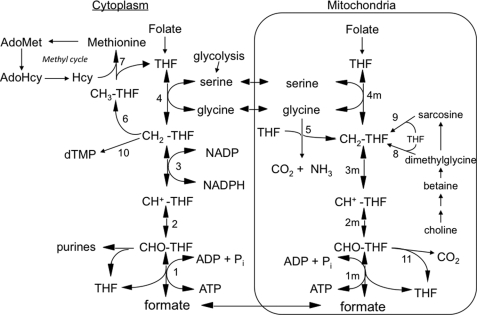
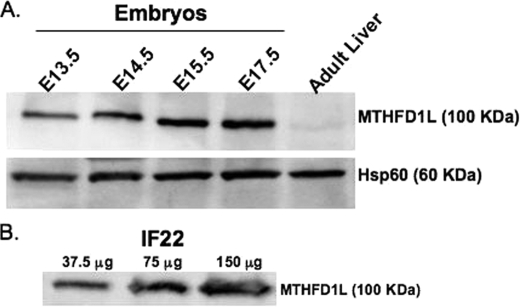
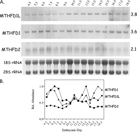
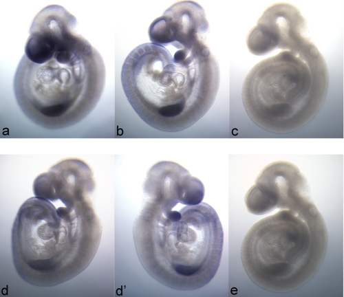
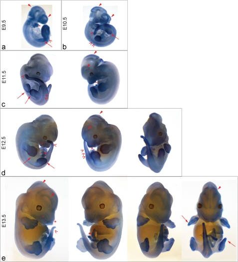
Mitochondrial folate-dependent one-carbon (1-C) metabolism converts 1-C donors such as serine and glycine to formate, which is exported and incorporated into the cytoplasmic tetrahydrofolate (THF) 1-C pool. Developing embryos depend on this mitochondrial pathway to provide 1-C units for cytoplasmic process such as de novo purine biosynthesis and the methyl cycle. This pathway is composed of sequential methylene-THF dehydrogenase, methenyl-THF cyclohydrolase, and 10-formyl-THF synthetase activities. In embryonic mitochondria, the bifunctional MTHFD2 enzyme catalyzes the dehydrogenase and cyclohydrolase reactions, but the enzyme responsible for the mitochondrial synthetase reaction has not been identified in embryos. A monofunctional 10-formyl-THF synthetase (MTHFD1L gene product) functions in adult mitochondria and is a likely candidate for the embryonic activity. Here we show that the MTHFD1L enzyme is present in mitochondria from normal embryonic tissues and embryonic fibroblast cell lines, and embryonic mitochondria possess the ability to synthesize formate from glycine. The MTHFD1L transcript was detected at all stages of mouse embryogenesis examined. In situ hybridizations showed that MTHFD1L was expressed ubiquitously throughout the embryo but with localized regions of higher expression. The spatial pattern of MTHFD1L expression was virtually indistinguishable from that of MTHFD2 and MTHFD1 (cytoplasmic C(1)-THF synthase) in embryonic day 9.5 mouse embryos, suggesting coordinated regulation. Finally, we show using stable isotope labeling that in an embryonic mouse cell line, greater than 75% of 1-C units entering the cytoplasmic methyl cycle are mitochondrially derived. Thus, a complete pathway of enzymes for supplying 1-C units from the mitochondria to the methyl cycle in embryonic tissues is established.


Figures

FIGURE 1.
FIGURE 1.
Embryonic mammalian one-carbon metabolism. Reactions 1–4 are in both the cytoplasmic and mitochondrial (m) compartments. Reactions 1–3, 10-formyl-THF synthetase, 5,10-methenyl-THF cyclohydrolase, and 5,10-methylene-THF dehydrogenase, respectively, are catalyzed by trifunctional C1-THF synthase in the cytoplasm (MTHFD1). In mammalian mitochondria, reaction 1m is catalyzed by monofunctional MTHFD1L, and reactions 2m and 3m are catalyzed by bifunctional MTHFD2. The other reactions are catalyzed by the following: serine hydroxymethyltransferase (reactions 4 and 4m), glycine cleavage system (reaction 5), 5,10-methylene-THF reductase (reaction 6), methionine synthase (reaction 7), dimethylglycine dehydrogenase (reaction 8), sarcosine dehydrogenase (reaction 9), thymidylate synthase (reaction 10), and 10-formyl-THF dehydrogenase (reaction 11; only the mitochondrial activity of this enzyme is shown, but it has been reported in both compartments in mammals). All of the reactions from choline to sarcosine are mitochondrial except the betaine to dimethylglycine conversion, which is cytoplasmic. Hcy, homocysteine; AdoHcy, S-adenosylhomocysteine.
FIGURE 2.
FIGURE 2.
Mitochondrial C1-THF synthase (MTHFD1L) is expressed in both embryos and MEFs. A, immunoblotting was performed on mitochondria isolated from E13.5 to E17.5 embryos and from adult liver. In each lane, 50 μg of total mitochondrial protein was loaded. The top panel shows MTHFD1L protein intensities. Hsp60 is a mitochondrial matrix marker used as a loading control. B, MTHFD1L protein was observed by immunoblot of increasing amounts of IF22 MEF whole cell extracts.
FIGURE 3.
FIGURE 3.
Temporal expression of MTHFD1L, MTHFD1, and MTHFD2 transcripts in mouse embryos. A, developmentally staged Northern blots were hybridized with probes to MTHFD1L, MTHFD1 and MTHFD2 transcripts. The age of the embryos from which the RNA was obtained is indicated above the blots in embryonic days. Approximate transcript sizes, in kilobases, is indicated on the right. The 18 and 28 S ribosomal RNAs from a photographic negative of the gel from which the blot was prepared are shown below the blots. These bands were used as loading controls. B, normalized relative transcript levels from each blot.
FIGURE 4.
FIGURE 4.
Spatial distribution of MTHFD1L and MTHFD2 expression in E9.5 mouse embryos. In situ hybridization patterns of MTHFD1L (top row) are identical for probes to the 5′ end (panel a) and 3′ end (panel b). MTHFD1L (panels a and b) and MTHFD2 (panels d and d′) demonstrate nearly identical expression profiles. The left and right sides of the same embryo stained for MTHFD2 are represented in panels d and d′, respectively. Panels c and e are E9.5 embryos stained with sense probes to the 5′ end of MTHFD1L and to MTHFD2, respectively. The images are all 40× magnification.
FIGURE 5.
FIGURE 5.
mRNA Expression of MTHFD1L in Mouse Embryos. In situ hybridization of MTHFD1L (5′ end probe) mRNA was performed on whole mouse embryos ranging from E9.5 to E13.5. Expression was observed throughout the embryos at all of these stages. MTHFD1L is especially prominent in the developing forebrain and midbrain (closed arrowheads); developing limb buds and digits (panels a–e, long arrows); somites, presomitic mesoderm, and developing tail (open arrowheads); and developing branchial arches and embryonic jaw (asterisks). Expression is observed in the developing vibrissae (open circles) at E12.5 (panel d) and E13.5 (panel e) and umbilicus (closed circle). The images were photographed at 40× magnification for E9.5 and E10.5 (panel a and b), 30× for E11.5 and E12.5 (panels c and d), and 12.5× for E13.5 embryos (panel e).

References

    1. Appling D. R. (1991) FASEB J. 5, 2645–2651 - PubMed
    1. Woeller C. F., Anderson D. D., Szebenyi D. M., Stover P. J. (2007) J. Biol. Chem. 282, 17623–17631 - PubMed
    1. Davis S. R., Stacpoole P. W., Williamson J., Kick L. S., Quinlivan E. P., Coats B. S., Shane B., Bailey L. B., Gregory J. F., 3rd (2004) Am. J. Physiol. Endocrinol. Metab. 286, E272–E279 - PubMed
    1. Fu T. F., Rife J. P., Schirch V. (2001) Arch. Biochem. Biophys. 393, 42–50 - PubMed
    1. Lamers Y., Williamson J., Theriaque D. W., Shuster J. J., Gilbert L. R., Keeling C., Stacpoole P. W., Gregory J. F., 3rd (2009) J. Nutr. 139, 666–671 - PMC - PubMed

Publication types

MeSH terms