. 2010 Jan 29;285(5):3245-52.
doi: 10.1074/jbc.M109.006320. Epub 2009 Nov 30.

Calcium binds to LipL32, a lipoprotein from pathogenic Leptospira, and modulates fibronectin binding

Affiliations

Calcium binds to LipL32, a lipoprotein from pathogenic Leptospira, and modulates fibronectin binding

Jung-Yu Tung et al. J Biol Chem. .

Abstract

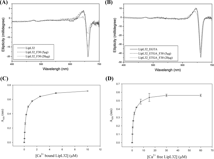
Tubulointerstitial nephritis is a cardinal renal manifestation of leptospirosis. LipL32, a major lipoprotein and a virulence factor, locates on the outer membrane of the pathogen Leptospira. It evades immune response by recognizing and adhering to extracellular matrix components of the host cell. The crystal structure of Ca(2+)-bound LipL32 was determined at 2.3 A resolution. LipL32 has a novel polyD sequence of seven aspartates that forms a continuous acidic surface patch for Ca(2+) binding. A significant conformational change was observed for the Ca(2+)-bound form of LipL32. Calcium binding to LipL32 was determined by isothermal titration calorimetry. The binding of fibronectin to LipL32 was observed by Stains-all CD and enzyme-linked immunosorbent assay experiments. The interaction between LipL32 and fibronectin might be associated with Ca(2+) binding. Based on the crystal structure of Ca(2+)-bound LipL32 and the Stains-all results, fibronectin probably binds near the polyD region on LipL32. Ca(2+) binding to LipL32 might be important for Leptospira to interact with the extracellular matrix of the host cell.


Figures

FIGURE 1.
FIGURE 1.
LipL32 sequence. The amino acid sequence and secondary structural elements of LipL32 are shown. The β-strands and α-helices are colored green and yellow, respectively. The polyD sequence (142DDDDDGDD149) is underlined. Asp113, Thr114, Asp145, Asp146, and Tyr159 are indicated (▴).
FIGURE 2.
FIGURE 2.
Ca2+-bound LipL32. A, the overall structure of Ca2+-bound LipL32. A ribbon drawing of four α-helices (yellow, labeled α1–α4) and 12 β-strands (green, labeled β1–β12) is shown. B, the Ca2+-binding site of LipL32. The Ca2+-coordinated residues Asp113, Thr114, Asp145, Asp146, and Tyr159 are shown by bonds and sticks and are colored according to Corey-Pauling-Koltun models. The hydrogen bonds are shown as dashed lines. The Ca2+ ion is shown as a yellow-colored sphere. C, the calcium binding affinity for LipL32 determined by ITC. Data for heat change obtained by calcium titration are shown in the upper panel. Data after peak integration (■) are shown in the lower panel, and the solid line represents the best fits to a single-site binding model.
FIGURE 3.
FIGURE 3.
Structural comparison between Ca2+-bound and Ca2+-free LipL32. A, the structural superimposition of Ca2+-bound LipL32 (this study) and Ca2+-free LipL32 (Protein Data Bank code 2ZZ8). The structural differences between Ca2+-bound and Ca2+-free LipL32 are colored green and blue, respectively. The Ca2+ ion is shown as a green-colored sphere. Strand β8 and loops β8β9 and β9β10 are labeled. The structural deviation of 10 Å on loop β8β9 (in His156 Cα) between Ca2+-bound and Ca2+-free LipL32 is labeled. The structural deviation of 18 Å on loop β9β10 (in Phe174 Cα) between Ca2+-bound and Ca2+-free LipL32 is also labeled. The missing region (Lys140–Asp146) of Ca2+-free LipL32 is represented by a dashed line. B, the major conformational change around loop β8β9. Asp113, Arg155, His156, Asn157, and Tyr198 are shown by bonds and sticks and are colored according to Corey-Pauling-Koltun models. The hydrogen bonds are shown as dashed lines. C, the possible collagen-binding site on LipL32. Leu53, Val54, Tyr62, Trp115, Arg117, and Tyr198 are shown by bonds and sticks.
FIGURE 4.
FIGURE 4.
Interaction between the fibronectin fragment F30 and LipL32. A and B, the Stains-all assay of the binding of F30 to Ca2+-bound and Ca2+-free LipL32, respectively. The CD spectra were recorded from 700 to 400 nm, and a J band at 660 nm is shown. C and D, the ELISA of the interaction between F30 and Ca2+-bound or Ca2+-free LipL32, respectively. Each point in C and D represents the mean absorbance of three independent experiments, and the error bars are the standard deviations for the mean of three independent data sets. The Kd values for F30 binding to LipL32 were calculated to be 0.29 ± 0.01 μm (Ca2+-bound LipL32) and 1.15 ± 0.06 μm (Ca2+-free LipL32).

Similar articles

Cited by

References

    1. Farr R. W. (1995) Clin. Infect. Dis. 21, 1–6 - PubMed
    1. Biegel E., Mortensen H. (1995) Ugeskr. Laeger 157, 153–157 - PubMed
    1. Bharti A. R., Nally J. E., Ricaldi J. N., Matthias M. A., Diaz M. M., Lovett M. A., Levett P. N., Gilman R. H., Willig M. R., Gotuzzo E., Vinetz J. M. (2003) Lancet 3, 757–771 - PubMed
    1. Yang C. W. (2007) Kidney Int. 72, 918–925 - PubMed
    1. Dolhnikoff M., Mauad T., Bethlem E. P., Carvalho C. R. (2007) Braz. J. Infect. Dis. 11, 142–148 - PubMed

Publication types

MeSH terms

Associated data

LinkOut - more resources