. 2009 Dec 15;23(24):2887-99.
doi: 10.1101/gad.1865909.

Quantitative proteomic analysis of purified yeast kinetochores identifies a PP1 regulatory subunit

Affiliations

Quantitative proteomic analysis of purified yeast kinetochores identifies a PP1 regulatory subunit

Bungo Akiyoshi et al. Genes Dev. .

Erratum in

Abstract

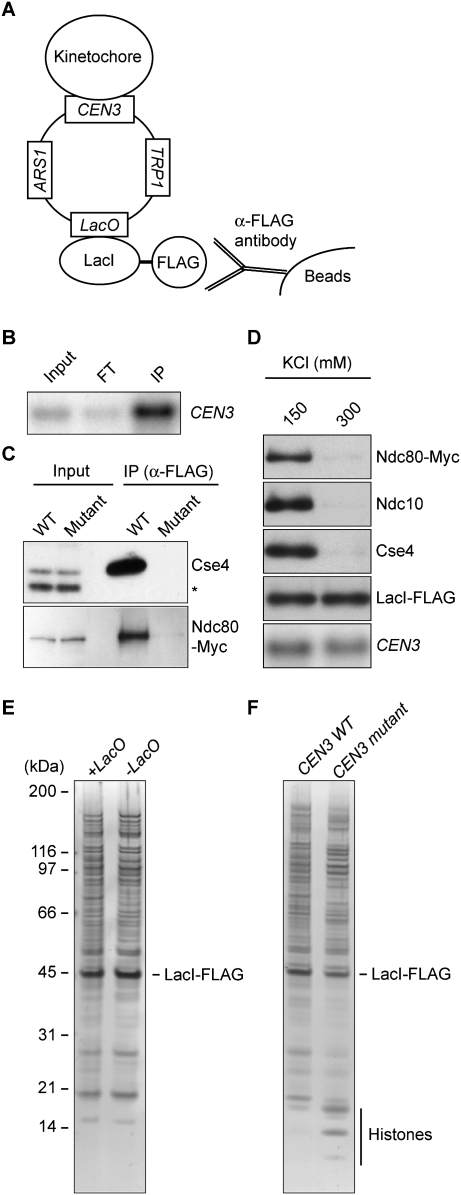
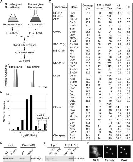
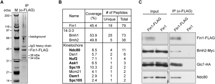
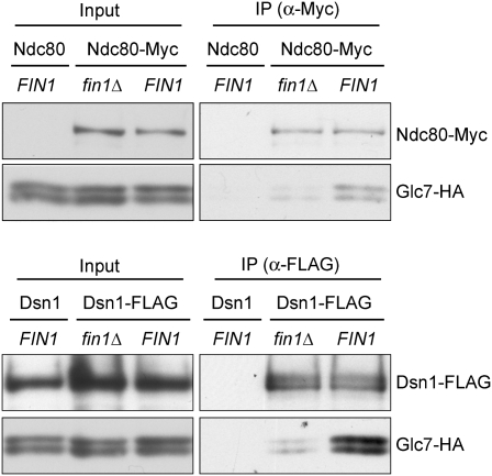
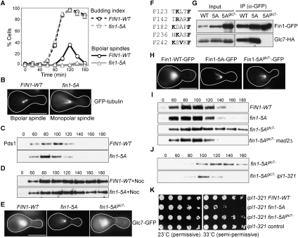
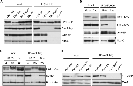
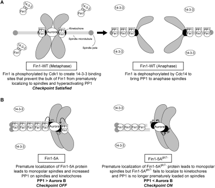
The kinetochore is a macromolecular complex that controls chromosome segregation and cell cycle progression. When sister kinetochores make bioriented attachments to microtubules from opposite poles, the spindle checkpoint is silenced. Biorientation and the spindle checkpoint are regulated by a balance between the Ipl1/Aurora B protein kinase and the opposing activity of protein phosphatase I (PP1). However, little is known about the regulation of PP1 localization and activity at the kinetochore. Here, we developed a method to purify centromere-bound kinetochores and used quantitative proteomics to identify the Fin1 protein as a PP1 regulatory subunit. The Fin1/PP1 complex is regulated by phosphorylation and 14-3-3 protein binding. When Fin1 is mislocalized, bipolar spindles fail to assemble but the spindle checkpoint is inappropriately silenced due to PP1 activity. These data suggest that Fin1 is a PP1 regulatory subunit whose spatial and temporal activity must be precisely controlled to ensure genomic stability.


Figures

Figure 1.
Figure 1.
Yeast kinetochores can be purified by isolating centromeric minichromosomes. (A) Minichromosome purification scheme. Centromeric minichromosomes containing LacO repeats are captured by LacI-Flag affinity purification. (B) Purified minichromosome DNA (SBY5218) was detected by Southern blot analysis using a probe specific to CEN3. Note that the immunoprecipitation (IP) lane contains 30 times more sample equivalent than input and flow-through (FT). (C) Kinetochore proteins remain associated with purified minichromosomes. Wild-type (SBY5218) or mutant (SBY5248) CEN3 minichromosomes were purified from strains expressing Ndc80-Myc. Immunoblots were performed using anti-Cse4 (asterisk indicates background band) and anti-Myc antibodies. (D) Kinetochores dissociate from CEN minichromosomes in the presence of 300 mM KCl. Minichromosomes (SBY5218) were washed with buffer containing 150 or 300 mM KCl. Purified samples were analyzed by immunoblots using antibodies to the indicated kinetochore proteins and by Southern blot analysis using a CEN3 probe to detect minichromosome DNA. (E) The LacI-Flag-purified samples have high levels of proteins that copurify independently of the lacO sequence. Proteins that copurified with minichromosomes that contained LacO repeats (SBY6107) or lacked the repeats (SBY6037) were analyzed by SDS-PAGE followed by silver staining to compare the sample purity. (F) The higher cellular copy number of mutant CEN minichromosomes increases the yield of mutant minichromosomes. Proteins that copurified with minichromosomes containing a wild-type (SBY6107) or a mutant (SBY6114) CEN were analyzed as in E. Note that histone bands were easily detected in the mutant CEN minichromosome purification.
Figure 2.
Figure 2.
Quantitative MS analysis identifies the Fin1 kinetochore protein. (A) Quantitative proteomics strategy. Centromeric minichromosomes (MCs) with LacO repeats (SBY6107) were grown in media containing isotopically heavy arginine and lysine (dark gray), while those without LacO (SBY6037) were grown in normal media (light gray). The graph shows that proteins specific to the heavy LacO-containing sample will be more abundant than proteins derived from the light sample. Background proteins will have abundance ratios that are similar or higher in the sample derived from the strain lacking the LacO sites. (B) Distribution of proteins detected by MS based on the enrichment ratio (H/L indicates heavy/light ratio). The dotted box indicates the proteins greater than fourfold-enriched in the heavy sample containing the LacO minichromosome. (C) List of kinetochore proteins with percentage of sequence coverage, number of unique and total peptides (N.D. indicates not detected), and H/L ratio with standard deviation (SD). (D) Fin1 associates with the centromere. Wild-type (SBY6010) or mutant (SBY6013) CEN3 minichromosomes were purified from cells that express Fin1-Myc and immunoblotted with anti-Myc antibodies. (E) The association of Fin1 with centromeric minichromosomes depends on kinetochore assembly. Wild-type CEN3 minichromosomes were purified from ndc10-1 (SBY6593) or NDC10 (SBY6594) strains and immunoblotted as in D. (F) Fin1 localizes to endogenous kinetochores during metaphase. Fin1-Myc cells (SBY8301) were arrested in metaphase by nocodazole treatment, and chromosome spreads were immunostained with anti-Cse4 and anti-Myc antibodies. Bar, 2 μm.
Figure 3.
Figure 3.
Fin1 associates with 14–3–3, outer kinetochore proteins, and PP1. (A,B) MS analysis identified Fin1-binding proteins. Fin1-Flag protein was purified from 5 L of asynchronously growing cells (SBY5962). (A) Sample was analyzed by SDS-PAGE followed by silver staining. Two proteins (36 kDa and 34 kDa) specifically copurified with Fin1-Flag. (B) Purified sample was analyzed by LC-MS/MS, and a summary is shown in the table. Outer kinetochore proteins are shown in bold. (C) Coimmunoprecipitation experiments confirmed that Fin1 specifically associates with 14–3–3, Ndc80, and the Glc7 phosphatase. Proteins were purified with anti-Flag antibodies from cells containing Bmh2-Myc and Glc7-HA that express either Fin1-Flag (SBY6368) or untagged Fin1 (SBY6370). Samples were analyzed by immunoblots with the corresponding antibodies.
Figure 4.
Figure 4.
Fin1 partially mediates the interaction between Glc7 and kinetochore proteins. (Top) Ndc80-Myc was immunoprecipitated from fin1Δ (SBY7895) and FIN1 (SBY7897) cells expressing Glc7-HA. Purified samples were analyzed by immunoblots using anti-Myc and anti-HA antibodies. Glc7-HA cells with untagged Ndc80 (SBY625) were used as negative control. (Bottom) Dsn1-Flag was purified with anti-Flag antibodies from fin1Δ (SBY7899) and FIN1 (SBY7900) cells expressing Glc7-HA. Purified samples were analyzed by immunoblots using anti-Flag and anti-HA antibodies. Note that the Dsn1-Flag band overlaps with a background signal in the input.
Figure 5.
Figure 5.
Fin1 regulates Glc7 and antagonizes the Ipl1 kinase. (A,B) Fin1-5A overexpression blocks bipolar spindle assembly. Cells with galactose-inducible FIN1-WT (SBY6825) or fin1-5A (SBY6809) expressing GFP-Tub1 were released from G1 into galactose and fixed every 20 min after release. Budding index and spindle morphology (bipolar or monopolar) were analyzed. Bar, 5 μm. (C) Fin1-5A cells do not activate the spindle checkpoint in response to spindle defects. Cells containing Pds1-Myc and pGAL-FIN1-WT (SBY6458) or pGAL-fin1-5A (SBY6459) were synchronized as in A. Lysates were prepared at the indicated time points and monitored for Pds1-Myc by immunoblot. (D) Cells expressing Fin1-5A activate the spindle checkpoint in response to microtubule depolymerization. The experiment in C was repeated by releasing cells into nocodazole. (E) Glc7 is mistargeted by Fin1-5A. Glc7-GFP localization was monitored in cells with galactose-inducible FIN1-WT (SBY6483), fin1-5A (SBY6484), or fin1-5Aglc7− (SBY6685) as in A. At 110 min after release, 89% of Fin1-5A cells showed Glc7 signal specifically on spindles and poles, while 100% of Fin1-WT and Fin1-5Aglc7 cells exhibited diffuse nuclear Glc7 staining in metaphase. (F) Fin1 possesses five potential Glc7-binding motifs. Residues that match the consensus PP1-binding sequence are highlighted in bold. (G) Fin1-5Aglc7 fails to bind Glc7. Asynchronously growing cells containing Glc7-HA and galactose-inducible FIN1-WT-GFP (SBY6497), fin1-5A-GFP (SBY6498), or fin1-5Aglc7−-GFP (SBY6662) were induced with galactose for 3 h. Fin1-GFP was immunoprecipitated with anti-GFP antibodies, and the samples were analyzed by immunoblots with anti-GFP and anti-HA antibodies. (H) Fin1-5Aglc7 prematurely localizes onto spindles. Cells expressing galactose-inducible FIN1-WT-GFP (SBY6915), fin1-5A-GFP (SBY6916), or fin1-5Aglc7−-GFP (SBY6917) were treated as in E. One-hundred percent of the metaphase cells expressing Fin1-5A-GFP or Fin1-5Aglc7-GFP cells showed premature spindle localization of Fin1 compared with 0% for Fin1-WT-GFP. (I) The spindle defect in Fin1-5Aglc7 cells triggers the spindle checkpoint. Cells containing Pds1-Myc and galactose-inducible FIN1-WT-GFP (SBY6573), fin1-5A-GFP (SBY6574), fin1-5Aglc7−-GFP (SBY6686), or fin1-5Aglc7−-GFP mad2Δ (SBY6824) were used to analyze checkpoint activation as in C. (J) Ipl1 activity is required for spindle checkpoint activation in Fin1-5Aglc7 cells. IPL1 (SBY6857) or ipl1-321 (SBY6858) cells containing Pds1-Myc and galactose-inducible Fin1-5Aglc7−-GFP were used to analyze checkpoint activation as in C, except that cells were released to 37°C. (K) Endogenous levels of Fin1-5A exhibit genetic interactions with an ipl1 mutant. Serial dilutions (fivefold) of ipl1-321 cells expressing FIN1-WT-GFP (SBY7352), fin1-5A-GFP (SBY7547), or fin1-5Aglc7−-GFP (SBY7548) from the endogenous promoter were plated at 23°C (permissive temperature) and 33°C (semipermissive temperature for ipl1-321). SBY7356 contains a control vector.
Figure 6.
Figure 6.
Regulation of Fin1 by phosphorylation, 14–3–3 proteins, and the PP1 phosphatase. (A) Phosphorylation is required for the Fin1 interaction with 14–3–3 proteins, and Glc7 binding is required for Fin1 to bind the Ndc80 kinetochore protein. Fin1-GFP proteins were immunoprecipitated with anti-GFP antibodies from cells containing Bmh2-Myc, Glc7-HA, and either FIN1-WT-GFP (SBY7609), fin1-5A-GFP (SBY7610), fin1-5Aglc7−-GFP (SBY7611), or fin1glc7−-GFP (SBY7627) expressed from the endogenous FIN1 promoter. The samples were analyzed by immunoblots with the indicated antibodies. (B) 14–3–3 proteins require phosphorylation to bind to Fin1. Cells containing Fin1-Flag, Bmh2-Myc, Glc7-HA ,and galactose-inducible nondegradable clb2 (SBY6452) were grown and then split into two cultures that were treated with either nocodazole (metaphase arrest) or galactose (late anaphase arrest) for 2.5 h. Fin1-Flag was immunoprecipitated, and samples were analyzed by immunoblots. (C) Glc7 is required for the Fin1-Ndc80 interaction. Fin1-Flag was purified from wild-type (SBY6373) or glc7-12 (SBY7841) strains that had been shifted to the restrictive temperature for 2.5 h, and copurifying proteins were analyzed by immunoblots. As a metaphase arrest control, wild-type cells treated with nocodazole were analyzed. (D) Glc7-binding mutants of Fin1 do not localize to kinetochores. Centromeric minichromosomes were purified from cells containing FIN1-WT-GFP (SBY7590), fin1-5A-GFP (SBY7591), fin1-5Aglc7−-GFP (SBY7592), or fin1glc7−-GFP (SBY7628) expressed from the endogenous FIN1 promoter. Samples were analyzed by immunoblots with anti-GFP and anti-Cse4 (loading control) antibodies.
Figure 7.
Figure 7.
Models for wild-type and mutant Fin1 protein function. (A) Wild-type Fin1 protein: (Left) In metaphase, Cdk1-dependent phosphorylation of Fin1 leads to 14–3–3 binding that prevents the bulk of Fin1 from prematurely localizing to spindles, thereby maintaining appropriate levels of the Aurora B kinase and PP1 phosphatase on kinetochores. There is also a pool of Fin1/PP1 complex at kinetochores that can oppose Aurora B. (Right) Once all kinetochores biorient, the checkpoint is satisfied and cells enter anaphase. During anaphase, the bulk of Fin1 is dephosphorylated by Cdc14 phosphatase to recruit Fin1 and PP1 to anaphase spindles. (B) Mutant Fin1 proteins: The premature localization of the Fin1-5A proteins (Fin1-5A and Fin1-5Aglc7) to microtubules results in monopolar spindles in a PP1-independent manner. (Left) The increased Fin1-5A protein on spindles and kinetochores inappropriately silences the spindle checkpoint. (Right) The Fin1-5Aglc7 fails to bind PP1 and kinetochores, so the spindle checkpoint remains active due to monopolar spindle formation.

Comment in

Similar articles

Cited by

References

    1. Akiyoshi B, Nelson CR, Ranish JA, Biggins S. Analysis of Ipl1-mediated phosphorylation of the Ndc80 kinetochore protein in Saccharomyces cerevisiae. Genetics. 2009 doi: 10.1534/genetics.109.109041. - DOI - PMC - PubMed
    1. Biggins S, Murray AW. The budding yeast protein kinase Ipl1/Aurora allows the absence of tension to activate the spindle checkpoint. Genes & Dev. 2001;15:3118–3129. - PMC - PubMed
    1. Biggins S, Severin FF, Bhalla N, Sassoon I, Hyman AA, Murray AW. The conserved protein kinase Ipl1 regulates microtubule binding to kinetochores in budding yeast. Genes & Dev. 1999;13:532–544. - PMC - PubMed
    1. Bloecher A, Tatchell K. Dynamic localization of protein phosphatase type 1 in the mitotic cell cycle of Saccharomyces cerevisiae. J Cell Biol. 2000;149:125–140. - PMC - PubMed
    1. Buvelot S, Tatsutani SY, Vermaak D, Biggins S. The budding yeast Ipl1/Aurora protein kinase regulates mitotic spindle disassembly. J Cell Biol. 2003;160:329–339. - PMC - PubMed

Publication types

MeSH terms