. 2010 Jan;16(1):1-9.
doi: 10.1261/rna.1791310. Epub 2009 Nov 30.

A structural analysis of the group II intron active site and implications for the spliceosome

Affiliations

A structural analysis of the group II intron active site and implications for the spliceosome

Kevin S Keating et al. RNA. 2010 Jan.

Abstract

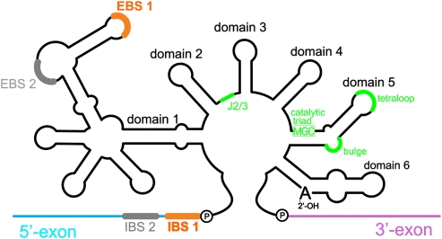
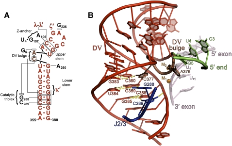
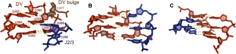
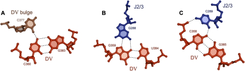
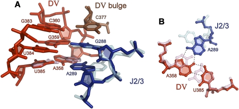
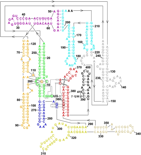
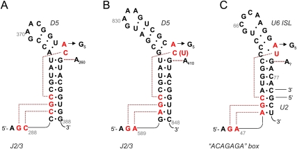
Group II introns are self-splicing, mobile genetic elements that have fundamentally influenced the organization of terrestrial genomes. These large ribozymes remain important for gene expression in almost all forms of bacteria and eukaryotes and they are believed to share a common ancestry with the eukaryotic spliceosome that is required for processing all nuclear pre-mRNAs. The three-dimensional structure of a group IIC intron was recently determined by X-ray crystallography, making it possible to visualize the active site and the elaborate network of tertiary interactions that stabilize the molecule. Here we describe the molecular features of the active site in detail and evaluate their correspondence with prior biochemical, genetic, and phylogenetic analyses on group II introns. In addition, we evaluate the structural significance of RNA motifs within the intron core, such as the major-groove triple helix and the domain 5 bulge. Having combined what is known about the group II intron core, we then compare it with known structural features of U6 snRNA in the eukaryotic spliceosome. This analysis leads to a set of predictions for the molecular structure of the spliceosomal active site.


Figures

FIGURE 1.
FIGURE 1.
Schematic of a generic group II intron secondary structure. The six intron domains radiate from a central wheel. The EBS sequences in D1 recognize the 5′-exon through base-pairing interactions with the IBS sequences (note that the O.i. intron does not have EBS2-IBS2). D5 and its conserved components are highlighted in green. Note that the MGC sequence in the D5 catalytic triad (M = A or C) forms a triple helix with sequences in J2/3, also indicated in green.
FIGURE 2.
FIGURE 2.
D5 structure showing interactions with other domains. (A) Secondary structure of D5. (B) Representation of the D5 tertiary structure. The catalytic bulge of D5 is shown in brown, while the rest of D5 is shown in red. The upstream terminus of the intron is shown in green and J2/3 is blue. Note that all group II intron structural analysis described in this paper was conducted on the refined model with pdb code 3EOH (Toor et al. 2008b). All interactions described and shown here are also present in the re-refined model with pdb code 3IGI (Toor et al. 2010).
FIGURE 3.
FIGURE 3.
Major-groove triplexes in RNA and DNA: (A) The catalytic triplex in D5. (B) The major-groove RNA triplex from telomerase RNA (Kim et al. 2008). (C) A typical DNA triplex consisting of three stacks (Rhee et al. 1999). Sugar puckers in the group II intron structure were determined using the phosphate–glycosidic bond perpendicular distance (Davis et al. 2007; Murray 2007), which measures the distance from the glycosidic bond to the 3′ phosphate. A distance of greater than 2.9 Å corresponds to a C3′ endo sugar pucker. This distance is 3.2 Å for C377, 3.5 Å for C289, and greater than 4.4 Å for all other triplex nucleotides, which indicates a C3′ endo pucker in all cases.
FIGURE 4.
FIGURE 4.
Base triples within the catalytic triplex. (A) Interaction between C377 of the DV bulge and C360–G383. (B) Interaction between G288 of J2/3 and C359–U384. (C) Interaction between C289 of J2/3 and C358–G385.
FIGURE 5.
FIGURE 5.
A model of the intron core containing the IIB consensus sequence for the catalytic triad (5′-AGC) and J2/3 (5′-GA). (A) The IIC structure of D5 and J2/3 are shown in transparent pink and cyan, respectively, while the IIB model of D5 and J2/3 are shown in red and dark blue, respectively. (B) A view from the top of the first triple interaction of the triplex. The A•A–U triple proposed for IIB introns is shown in bold, superimposed on the transparent C•C–G triple observed in the IIC crystal structure. The IIB model was constructed starting from the IIC structure. Nucleotide bases were mutated and manually repositioned using Coot (Emsley and Cowtan 2004). Energy minimization was then carried out using CNS (Brünger et al. 1998). Minimization constraints for the A•A interaction were derived from structures found in the base pair isostericity database (Leontis et al. 2002).
FIGURE 6.
FIGURE 6.
Revised secondary structure of the O.i. intron. Domains I(i) and I(ii) are shown in green, IA and IB in purple, IC in orange, ID1 in gray, ID2 in cyan, DII in blue, DIII in yellow, DIV in beige, DV in red, and DVI (which was not visualized in the crystal structure) in black. Tertiary base-pairing (shown as circles), base-triple (shown as squares), and base-stacking interactions (shown as rectangles) are displayed only for the core of the intron.
FIGURE 7.
FIGURE 7.
Tertiary interactions involving D5 and, hypothetically, U6. (A) Interactions observed crystallographically for the O.i. group IIC intron. (B) Cognate interactions inferred for the ai5γ group IIB intron (B). These are readily modeled into the O.i. crystal structure (see Fig. 5), and they are consistent with chemogenetic experiments, phylogenetic data, and crosslinking results. (C) Cognate interactions are inferred for the eukaryotic spliceosome. Note that U6 is schematically represented in the same manner as is shown in Figure 5d by Sashital et al. (2004). This secondary structural representation is a U6 helix 1b model for the human spliceosomal active site (Madhani and Guthrie 1992) in which most of the U6 ISL is also present. This form has been proposed to be operative during the second step of splicing (Sun and Manley 1995; Sashital et al. 2004), but the interactions shown and proposed in this paper are equally consistent with the U6 ISL model for U6 (Sun and Manley 1995), which has been proposed to be operative during the first step of splicing (Sun and Manley 1995; Sashital et al. 2004). Based on an alignment composed of the 203 seed sequences from the Rfam database (http://rfam.janelia.org/cgi-bin/getdesc?name=U6) (Griffiths-Jones et al. 2005; Davila Lopez et al. 2008), the following levels of conservation are seen for nucleotides involved in the proposed interactions: G45 (98%), A46 (99%), A53 (98%), G54 (99%), C55 (100%), A73 (91%), U74 (95%), and G75 (99%).

Similar articles

Cited by

References

    1. Abramovitz DL, Friedman RA, Pyle AM. Catalytic role of 2′-hydroxyl groups within a group II intron active site. Science. 1996;271:1410–1413. - PubMed
    1. Barre FX, Ait-Si-Ali S, Giovannangeli C, Luis R, Robin P, Pritchard LL, Helene C, Harel-Bellan A. Unambiguous demonstration of triple-helix-directed gene modification. Proc Natl Acad Sci. 2000;97:3084–3088. - PMC - PubMed
    1. Blad H, Reiter NJ, Abildgaard F, Markley JL, Butcher SE. Dynamics and metal ion binding in the U6 RNA intramolecular stem–loop as analyzed by NMR. J Mol Biol. 2005;353:540–555. - PubMed
    1. Boeke JD. The unusual phylogenetic distribution of retrotransposons: A hypothesis. Genome Res. 2003;13:1975–1983. - PubMed
    1. Boulanger SC, Belcher SM, Schmidt U, Dib-Hajj SD, Schmidt T, Perlman PS. Studies of point mutants define three essential paired nucleotides in the domain 5 substructure of a group II intron. Mol Cell Biol. 1995;15:4479–4488. - PMC - PubMed

Publication types

LinkOut - more resources