. 2010 Jan;16(1):186-96.
doi: 10.1261/rna.1847310. Epub 2009 Nov 30.

Thermodynamic analysis of ligand binding and ligand binding-induced tertiary structure formation by the thiamine pyrophosphate riboswitch

Affiliations

Thermodynamic analysis of ligand binding and ligand binding-induced tertiary structure formation by the thiamine pyrophosphate riboswitch

Nadia Kulshina et al. RNA. 2010 Jan.

Abstract

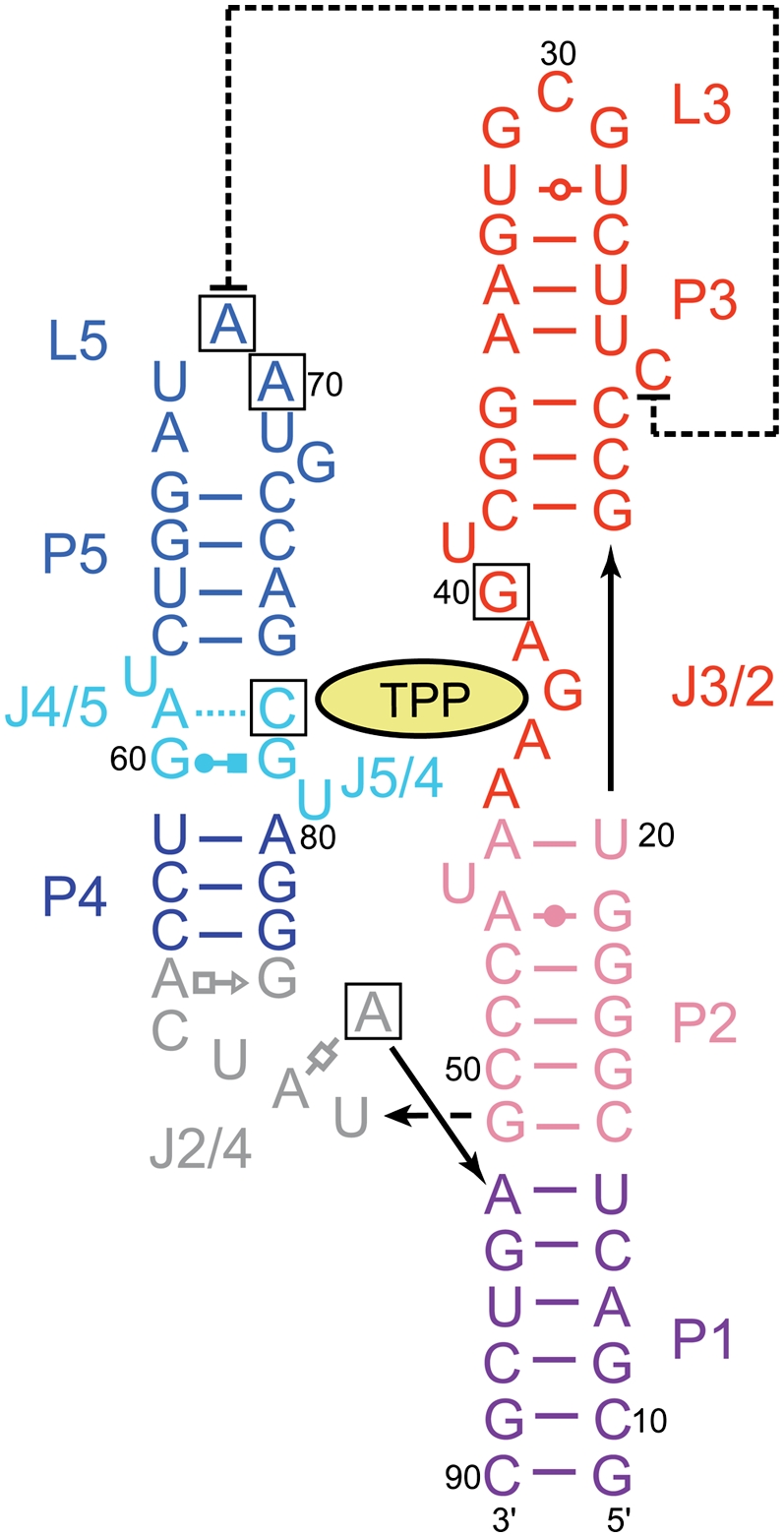
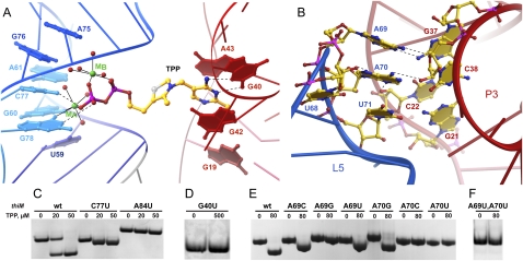
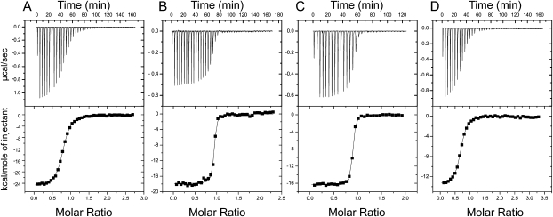
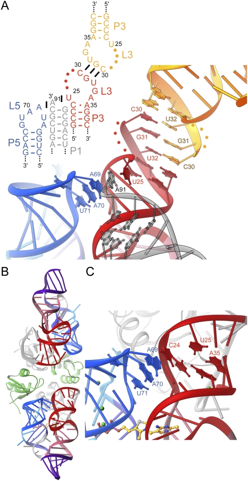
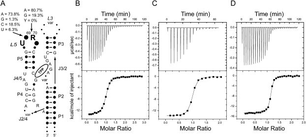
The thi-box riboswitch regulates gene expression in response to the intracellular concentration of thiamine pyrophosphate (TPP) in archaea, bacteria, and eukarya. To complement previous biochemical, genetic, and structural studies of this phylogenetically widespread RNA domain, we have characterized its interaction with TPP by isothermal titration calorimetry. This shows that TPP binding is highly dependent on Mg(2+) concentration. The dissociation constant decreases from approximately 200 nM at 0.5 mM Mg(2+) concentration to approximately 9 nM at 2.5 mM Mg(2+) concentration. Binding is enthalpically driven, but the unfavorable entropy of binding decreases as Mg(2+) concentration rises, suggesting that divalent cations serve to pre-organize the RNA. Mutagenesis, biochemical analysis, and a new crystal structure of the riboswitch suggest that a critical element that participates in organizing the riboswitch structure is the tertiary interaction formed between the P3 and L5 regions. This tertiary contact is distant from the TPP binding site, but calorimetric analysis reveals that even subtle mutations in L5 can have readily detectable effects on TPP binding. The thermodynamic signatures of these mutations, namely decreased favorable enthalpy of binding and small effects on entropy of binding, are consistent with the P3-L5 association contributing allosterically to TPP-induced compaction of the RNA.


Figures

FIGURE 1.
FIGURE 1.
Sequence and secondary structure of the E. coli thiM riboswitch, based on the structure given by Serganov et al. (2006). (Black boxes) Site-directed mutations examined in this study, (lines with arrowheads) connectivity. Noncanonical pairs are denoted with Leontis–Westhof (Leontis and Westhof 2001) symbols. (Dashed line) Tertiary stacking interaction.
FIGURE 2.
FIGURE 2.
Importance for TPP binding of structural features of the thi-box riboswitch. (A) Detail of the TPP binding pocket, with magnesium ion-dependent recognition of the pyrophosphate binding (left) and recognition of the aminopyrimidine by base pairing with G40 and stacking interactions. (Dashed lines) Hydrogen bonds, (solid lines) inner-sphere coordination. (B) P3–L5 interactions. (C) EMSA analysis shows that the mutation C77U is permissive for binding, whereas mutations A84U and G40U abolish binding (D). (E,F) EMSA analysis of mutants in positions 69 and 70 shows that only A69C, A69U, and A70G are permissive of binding. RNA concentration was 40 μM in CF.
FIGURE 3.
FIGURE 3.
ITC analysis of TPP and TMP binding by the wild-type thiM thi-box aptamer domain. Representative TPP titrations at 0.5 mM (A), 2.5 mM (B), and 10.0 mM (C) Mg2+ concentration. (D) TMP titration at 10 mM Mg2+ concentration.
FIGURE 4.
FIGURE 4.
Structural plasticity of P3. (A) Schematic secondary structure and cartoon representation of the P3–L5 region of the riboswitch in its crystalline context, in the structure given by Edwards and Ferré-D'Amaré (2006). Interactions formed with symmetry-related molecules in the crystal (gray and yellow) are shown. Note intercalation of A91 from a symmetry-related molecule between U25 and A69. (B) Cartoon representation of the new E. coli thiM aptamer domain crystal structure. The two RNA molecules in the asymmetric unit are colored as in Figure 1. (Gray) Cognate U1A-RBDs, (green) two additional U1A-RBDs that do not make specific RNA-binding interactions. (C) Detail of P3–L5 interaction in the new crystal structure. Note extrusion of C24 from P3, and its stacking on A69.
FIGURE 5.
FIGURE 5.
ITC analysis of the L3–P5 interaction. (A) Schematic representation of phylogenetic sequence conservation of the thi-box riboswitch aptamer domain. (Solid circles, “var”) Variable positions. A summary of the analysis of 500 sequences from the Rfam database (Gardner et al. 2009) is given for positions 69 and 70. (B) Representative ITC experiment analyzing binding of TPP to E. coli thiM aptamer domain mutant A69C. (C) TPP titration of mutant A69U. (D) TPP titration of mutant A70G. All experiments were performed in the presence of 10 mM Mg2+.

Similar articles

Cited by

References

    1. Alam S, Grum-Tokars V, Krucinska J, Kundracik ML, Wedekind JE. Conformational heterogeneity at position U37 of an all-RNA hairpin ribozyme with implications for metal binding and the catalytic structure of the S-turn. Biochemistry. 2005;44:14396–14408. - PubMed
    1. Batey RT, Gilbert SD, Montange RK. Structure of a natural guanine-responsive riboswitch complexed with the metabolite hypoxanthine. Nature. 2004;432:411–415. - PubMed
    1. Bocobza S, Adato A, Mandel T, Shapira M, Nudler E, Aharoni A. Riboswitch-dependent gene regulation and its evolution in the plant kingdom. Genes & Dev. 2007;21:2874–2879. - PMC - PubMed
    1. Brünger AT, Adams PD, Clore GM, Gros P, Grosse-Kunstleve RW, Jiang J-S, Kuszewski J, Nilges M, Pannu NS, Read RJ, et al. Crystallography and NMR system: A new software system for macromolecular structure determination. Acta Crystallogr D Biol Crystallogr. 1998;54:905–921. - PubMed
    1. Canny MD, Jucker FM, Kellogg E, Khvorova A, Jayasena SD, Pardi A. Fast cleavage kinetics of a natural hammerhead ribozyme. J Am Chem Soc. 2004;126:10848–10849. - PubMed

Publication types

MeSH terms

LinkOut - more resources