. 2009 Nov;21(11):3591-609.
doi: 10.1105/tpc.109.065557. Epub 2009 Nov 30.

Arabidopsis N-MYC DOWNREGULATED-LIKE1, a positive regulator of auxin transport in a G protein-mediated pathway

Affiliations

Arabidopsis N-MYC DOWNREGULATED-LIKE1, a positive regulator of auxin transport in a G protein-mediated pathway

Yashwanti Mudgil et al. Plant Cell. 2009 Nov.

Abstract

Root architecture results from coordinated cell division and expansion in spatially distinct cells of the root and is established and maintained by gradients of auxin and nutrients such as sugars. Auxin is transported acropetally through the root within the central stele and then, upon reaching the root apex, auxin is transported basipetally through the outer cortical and epidermal cells. The two Gbetagamma dimers of the Arabidopsis thaliana heterotrimeric G protein complex are differentially localized to the central and cortical tissues of the Arabidopsis roots. A null mutation in either the single beta (AGB1) or the two gamma (AGG1 and AGG2) subunits confers phenotypes that disrupt the proper architecture of Arabidopsis roots and are consistent with altered auxin transport. Here, we describe an evolutionarily conserved interaction between AGB1/AGG dimers and a protein designated N-MYC DOWNREGULATED-LIKE1 (NDL1). The Arabidopsis genome encodes two homologs of NDL1 (NDL2 and NDL3), which also interact with AGB1/AGG1 and AGB1/AGG2 dimers. We show that NDL proteins act in a signaling pathway that modulates root auxin transport and auxin gradients in part by affecting the levels of at least two auxin transport facilitators. Reduction of NDL family gene expression and overexpression of NDL1 alter root architecture, auxin transport, and auxin maxima. AGB1, auxin, and sugars are required for NDL1 protein stability in regions of the root where auxin gradients are established; thus, the signaling mechanism contains feedback loops.


Figures

Figure 1.
Figure 1.
Protein Interaction, NDL Gene Family, and Putative Orthologs, and NDL and Similar Protein Structure. (A) Yeast strain AH109 was transformed with two plasmids: the pBridge (DNA-BD) vector containing AGB1/AGG1 or AGB1/AGG2 and the Gal4ACTD conjugated construct having NDL1, NDL2, NDL3, or NDRG1 (mouse NDRG1). The cells were grown on SC medium lacking Trp, Leu, His, and adenine (-W-L-H-Ade) or SC-W-L-H medium minus Met to repress AGG expression (-W-L-M-H). All three NDL proteins interacted with AGB1+AGG2/or AGG1 in a yeast three-hybrid assay. Interactions were scored on the basis of activation of the HIS3 reporter gene, X-gal staining, and presence or absence of Met. Single-domain controls: strain AH109 was transformed with single-domain plasmids alone and grown on selection. Using the same approach, the interactions of the prey set were also tested against the C-terminal cytoplasmic domain (C4) of Arabidopsis RGS1. (B) AGB1 and NDL1 interaction in planta. After 22 h of Agrobacterium tumefaciens–mediated transient expression of FLAG:AGB1 (F-AGB1) and CFP:NDL1 (C-NDL1) in wild-type N. benthamiana leaves, total protein was isolated and was immunoprecipitated with anti-FLAG (for AGB1) and anti-GFP (for NDL1) antibodies. Immunoprecipitated (IP) proteins were detected by immunoblotting with the indicated antibody (IB). Lane 1, IP of NDL1 with anti GFP antibodies; lane 2, IP of AGB1 with anti FLAG antibodies; lane 3, wild-type N. benthamiana extract (N.B) IP with anti FLAG. NDL1 coimmunoprecipitates with AGB1, and the reciprocal coimmunoprecipitation also occurred (lanes 1 and 2). Arrowheads indicate the position of NDL1 and AGB1. Brackets highlight the absence of AGB1 in the control. Protein masses are indicated at the left side of the immunoblots (in kilodaltons). (C) Phylogenetic tree of NDR proteins: all plant NDR homologs form a separate group in the unrooted tree and are highlighted by the red circle (X.l, Xenopus laevis; H.s, Homo sapiens; M.m, Mus musculus; A.g, Anopheles gambiae; H.a, Helianthus annuus; O.t, Ostreococcus tauri; O.s, Oryza sativa; D.m, Drosophila melanogaster; C.e, Caenorhabditis elegans). The phylogenetic tree was built as described in Methods. (D) NDL proteins are highly similar with conserved domains: amino acid alignment of the three Arabidopsis NDL proteins shows that all three NDL proteins contain the conserved NDR domain (red), an overlapping α/β hydrolase fold (underlined), a conserved Asp (boxed), a conserved hydrophobic patch (green), and catalytic triad residues marked with arrowheads. (E) Atomic model of NDL1: Surface representation (light green) of the active site pocket in NDL1 with overlaying flap (purple). Conserved D (TYPD) in pocket is colored red. (F) Surface representation (violet) of the active site pocket in the 2PU5 template with the overlying flap in yellow. Active site residues (S112, D237, and H265) are colored red.
Figure 2.
Figure 2.
NDL1 Tissue and Organ Localization. (A) to (S) In situ localization of NDL protein was indirectly determined using translational NDL-GUS fusion lines (T3). Bars = 50 μm in (A) to (G) and (I), 20 μm in (L) to (S), and 10 μm in (H), (J), and (K). (P) to (S) have the same magnification for direct comparison. (A) One- to two-day-old seedlings with emerging radicle. Arrow indicates staining at the root tip. (B) Two- to three-day-old, light-grown seedling. Arrow indicates staining at the root-shoot junction. (C) Ten-day-old, light-grown plant. (D) Stipules at the leaf base. (E) Mature true leaf, with red arrows showing localization at the base of trichomes. (F) Mature flower. (G) Stamen. (H) GUS-stained and fixed mature pollen grain. (I) Germinated GUS-stained and fixed pollen grain. (J) Head of the germinated GUS-stained and fixed pollen grain at higher magnification. (K) Tip of the germinated GUS-stained and fixed pollen grain at higher magnification. (L) Cross section of the primary root tip region; red arrows indicate comparatively deep staining at the endodermal layer. (M) Cross section of primary root around basal meristem. (N) Longitudinal section of primary root showing apical and basal meristematic zones. (O) Longitudinal section of primary root from the basal meristem region (marked by red arrows in [N]) at higher magnification. (P) Stage I of lateral root primordium development; red arrows point toward individual cells in layer. (Q) Stage II of lateral root primordium development. (R) Stage III of lateral root primordium development. (S) Stage IV of lateral root primordium development.
Figure 3.
Figure 3.
NDL1 Subcellular Localization. (A) Laser scanning confocal micrograph showing cytoplasmic localization of C-terminally GFP-tagged NDL1 stably expressed in Arabidopsis root epidermal cells under the transcriptional control of the NDL1 promoter. (B) Corresponding differential interference contrast image to the image shown in (A). (C) Control epidermal cell not expressing a GFP-tagged NDL1. (D) Corresponding differenetial interference contrast image shown in (C). Bars = 10 μm in (A) to (D). (E) and (F) Spinning disc confocal micrographs showing cytoplasmic localization of N-terminally (E) and N- plus C-terminally GFP-tagged (F) 35S-NDL1 transiently expressed in N. benthamiana. Bars = 20 μm.
Figure 4.
Figure 4.
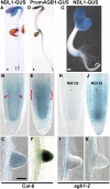
In Vivo Localization Pattern of NDL1 Protein in the Root in the Presence and Absence of AGB1. (A) NDL-GUS staining pattern in the wild-type seedling. Bracket indicates area shown in (B). (B) NDL1-GUS staining pattern in the wild-type root tip. (C) Lateral root staining pattern of NDL1 in wild-type (Col-0) background. (D) Transgenic plant expressing transcriptional fusion of the AGB1 promoter with GUS. (E) AGB1 expression in the root tip. (F) Lateral root primordium expression of AGB1. (G) NDL1-GUS staining pattern in the agb1-2 background. (H) Lateral root staining pattern of the steady state level of NDL1 in the agb1-2 background. NDL1 was not detectable (−) around the RAM in primary and lateral roots in the absence of AGB1. Compare the bracketed region of (A) to the bracketed region of (G), and (B) and (H) for an enlarged view. (J) and (K) MG132 treatment (100 μM for 4 h) of 4-d-old seedlings resulted in reappearance of NDL protein (+) in the primary and lateral roots in the agb1-2 background. Bars = 50 μm; the middle and bottom rows have the same magnification. Fifteen independent T1 GUS-positive, 3-d-old, light-grown seedlings were analyzed. Further expression analysis in lateral roots was performed with four independent T2 lines.
Figure 5.
Figure 5.
Effect on Root Length and Lateral Root Density by Altering NDL Levels in the Presence and Absence of AGB1. All experiments were repeated three times using 10 to 15 seedlings for each genotype in each trial. (A) Root length (mm) of 9- to 10-d-old, short-day-grown seedlings (8:16, light:dark). The genotypes (described in the text) are indicated below (B). (B) Lateral root density (primordia and emergent roots per centimeter of primary root length) for roots described in (A). (C) Number of lateral roots with (black bars) and without (open bars) induction by 0.1 μM napthalene-l-acetic acid. (D) mRNA quantification of all the genotypes used was performed by qRT-PCR using gene-specific primers for NDL1, normalized to the ACTIN2 transcript level. Expression level of NDL2 and NDL3 in ndlM1 and ndlM2 lines is shown as an inset. Error bars represent se. Student's t test results are based on differences between the wild type and the indicated genotype shown as asterisks: **, P < 0.05; ***, P < 0.005.
Figure 6.
Figure 6.
Relative Auxin Transport and Expression Level of PIN2 and AUX1 in Various G protein and NDL Genotypes. (A) Basipetal auxin transport measured by applying [3H]-IAA to the root apex and root-shoot junction as described in Methods. (B) Acropetal transport measured as described in Methods. For both basipetal and acropetal transport, means ± se are shown. The means are based on at least five independent trials, each involving >10 roots per genotype. Student's t test analysis based on differences between the wild type and the indicated genotype are indicated by asterisks above the bars: ***, P < 0.001; **, P < 0.05. (C) qRT-PCR showing relative expression levels of PIN2 and AUX1 upon downregulation and overexpression of NDL1 and in agb1-2 and rgs1-2 mutants. Data represent means ± se of three replicates; similar results were obtained in three independent biological replicates. Student's t analysis based on differences between the wild type and the indicated genotypes are indicated by asterisks above the bars: ***, P < 0.0001. (D) to (F) Effect of auxin application on NDL1 protein levels in the wild type (D) and agb1-2 background (E) and on AGB1 expression levels (F). NDL1-GUS translational fusion and ProAGB1-GUS lines were treated with 1 μM IAA, for 12 to 14 h, followed by GUS staining. Auxin decreased NDL1 steady state level and auxin increased AGB1 expression compared with the untreated controls (c.f. Figures 4B, 4E, and 4H). Bar = 50 μm; each panel is equivalent in magnification.
Figure 7.
Figure 7.
Auxin Maxima in the Wild Type and in Lines with Reduced Expression of NDL Genes. (A) to (D) Spatial pattern of the auxin reporter, DR5-GUS, in the wild-type background. Black arrows indicate lateral root primordia. (B) to (D) Higher magnification of auxin maxima observed at the apical meristem (B), root (C), and root tip (D). (E) DR5-GUS pattern in lines with reduced expression of the NDL gene family using miRNA in the reporter background. Loss of NDL proteins decreased the number and intensity of auxin maxima. (F) Detailed view of the tip still showing deep staining pattern in (E). (G) DR5-GUS in the wild-type background treated with 0.1 μM IAA for 14 h. (H) to (J) As for (B) to (D) except for the root shown in (G). (K) DR5-GUS expression patterns in the silenced NDL background with IAA induction. (L) to (N) As for (B) to (D) except of the root shown in (K). Genotypes are indicated. Bars = 50 μm.
Figure 8.
Figure 8.
Steady State Levels of NDL1-GUS and AGB1 Expression in Response to Various Sugar Treatments in the Presence and Absence of AGB1. (A) to (F) Five-day-old dark-grown seedlings were treated with 100 mM sucrose or d-glucose for 8 h, followed by X-gluc staining. The optimal time and dose was predetermined for NDL-GUS (see Supplemental Figure 8 online). (A) and (B) NDL-GUS level in response to d-glucose (A) or sucrose (B). (C) and (D) ProAGB1-GUS expression in response to d-glucose (C) or sucrose (D). (E) and (F) NDL steady state levels in the agb1-2 background in the presence of d-glucose (E) or sucrose (F). (G) to (I) Control, dark-grown seedlings on NDL-GUS seedlings on half-strength MS medium without sugars (G), with l-glucose (H), and with sorbitol (I). These controls showed no staining for NDL-GUS. For the untreated control of ProAGB-GUS and agb1-2, see Figures 4E and 4H, respectively. (A) to (I) have same scale bar as in (G). (J) and (K) Sugar-induced lateral root formation in the absence (J) and presence (K) of NPA in various G protein and NDL genotypes. Student's t test analysis based on differences between sugar treatment of the wild type and the indicated genotypes are indicated by asterisks above the se: ***, P < 0.001; **, P < 0.05. Sugar treatments are compared to control in the wild type or controls among all genotypes. These experiments were repeated three times, and the same pattern of lateral root formation was observed. For each experiment, 15 to 20 seedlings were counted. (K) Same as (J) except 5 μM NPA was included. (L) to (O) Three-day-old, dark-grown seedlings were treated with 300 mM sucrose for 12 h ([N] and [O]) and compared with the untreated control ([L] and [M]). Red arrows indicate increased areas of GUS staining. (M) and (O) represent high magnification of (L) and (N), respectively. Bars = 50 μm.
Figure 9.
Figure 9.
Proposed Physical Relationship of NDL in the G Protein Complex and a Model of the Mode of Action of NDL. (A) Physical interaction model. NDL1 is shown as part of the G protein–coupled pathway on the membrane. Interactions that have been shown here are between the AGB1 and RGS1 with NDL proteins. Previous work described in the text supports interactions between RGS1 and GPA1 and between GPA1 and AGB1 in Arabidopsis. (B) Genetic and biochemical interaction model. Epistasis analysis predicts that AGB1 and NDL proteins act, at least in part, via independent parallel pathways. The genetic data are also consistent with AGB1 and NDL1 acting in a complex where NDL1 is a positive and AGB1 is a negative regulator of lateral root formation, but the mechanism is unclear as represented by the bracket. NDL1 and AGB1 regulate auxin-induced lateral root formation via their effect on auxin transport. NDL1 promotes the flux of the basipetal stream of auxin transport and hence on lateral root initiation. AGB1 has the opposite action. AGB1, sugars, and auxin operate on NDL in the feedback loops indicated by the wavy lines. The scheme does not illustrate the redundant nature of the three NDL proteins. L.R., lateral root.

Similar articles

Cited by

References

    1. Akgoz, M., Azpiazu, I., Kalyanaraman, V., and Gautam, N. (2002). Role of the G protein gamma subunit in beta gamma complex modulation of phospholipase Cβ function. J. Biol. Chem. 277 19573–19578. - PubMed
    1. Bainbridge, K., Guyomarc'h, S., Bayer, E., Swarup, R., Bennett, M., Mandel, T., and Kuhlemeier, C. (2008). Auxin influx carriers stabilize phyllotactic patterning. Genes Dev. 22 810–823. - PMC - PubMed
    1. Beemster, G.T., and Baskin, T.I. (1998). Analysis of cell division and elongation underlying the developmental acceleration of root growth in Arabidopsis thaliana. Plant Physiol. 116 1515–1526. - PMC - PubMed
    1. Benjamins, R., Malenica, N., and Luschnig, C. (2005). Regulating the regulator: the control of auxin transport. Bioessays 27 1246–1255. - PubMed
    1. Benjamins, R., and Scheres, B. (2008). Auxin: The looping star in plant development. Annu. Rev. Plant Biol. 59 443–465. - PubMed

Publication types

MeSH terms

Substances