. 2010 Jan;176(1):51-63.
doi: 10.2353/ajpath.2010.080843. Epub 2009 Nov 30.

Inducible nephrin transgene expression in podocytes rescues nephrin-deficient mice from perinatal death

Affiliations

Inducible nephrin transgene expression in podocytes rescues nephrin-deficient mice from perinatal death

Juuso Juhila et al. Am J Pathol. 2010 Jan.

Abstract

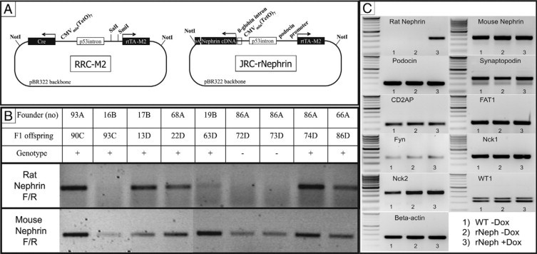
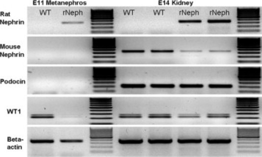
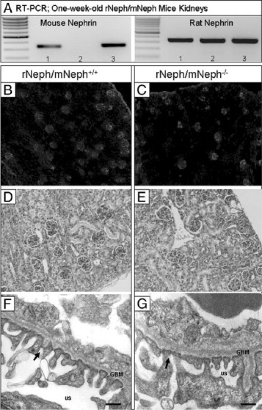
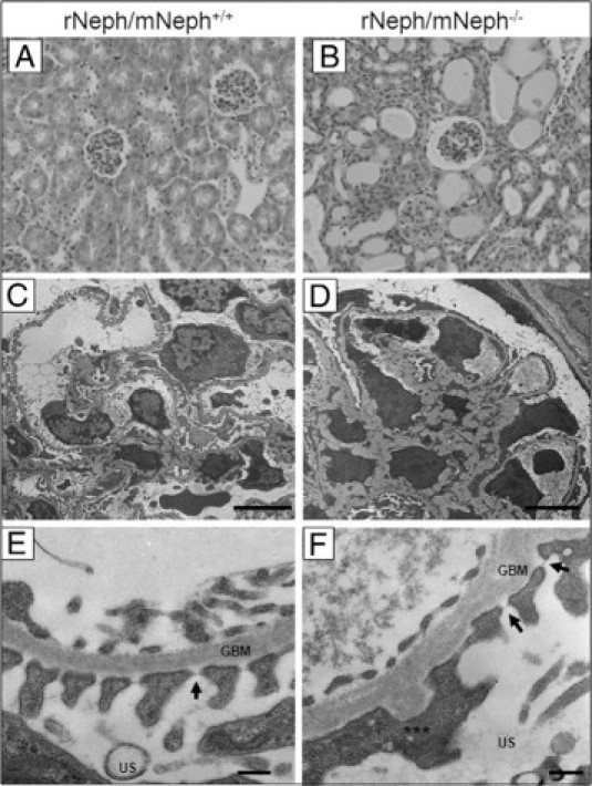
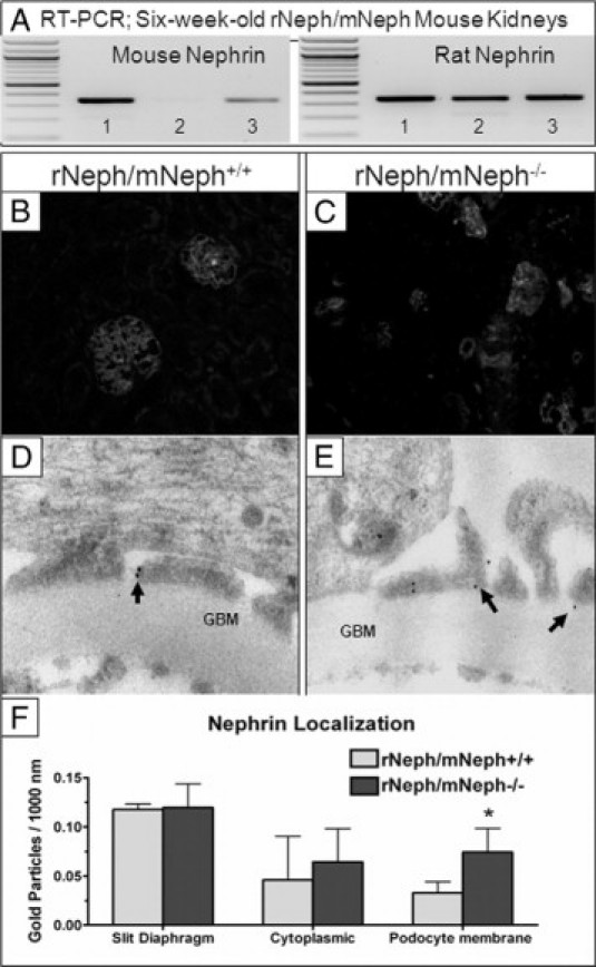
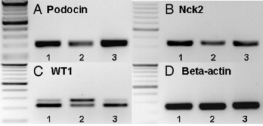
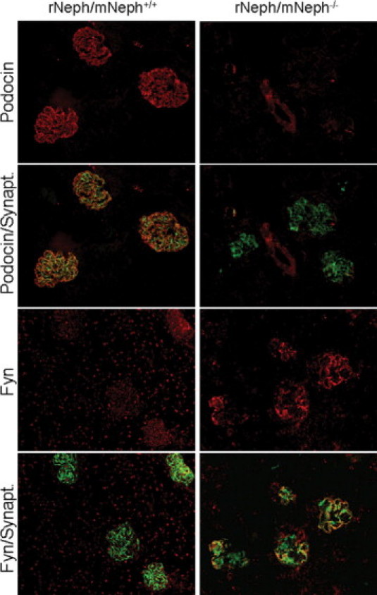
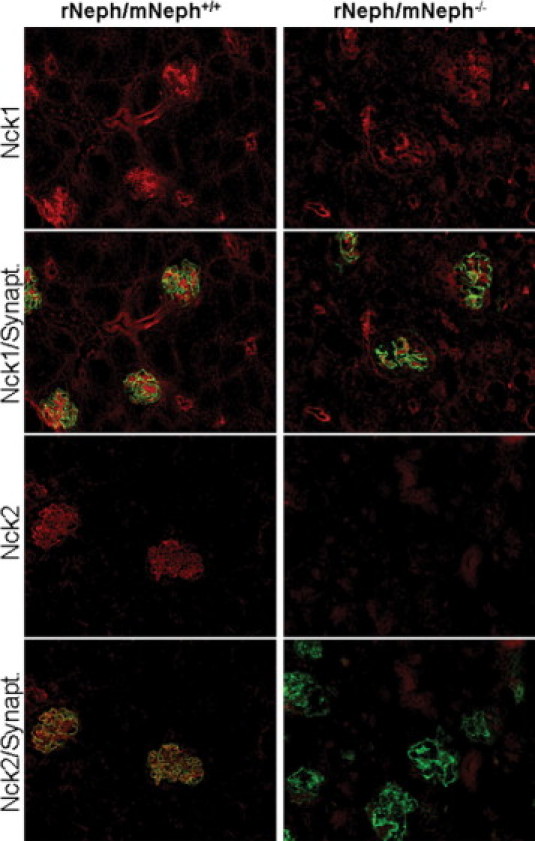
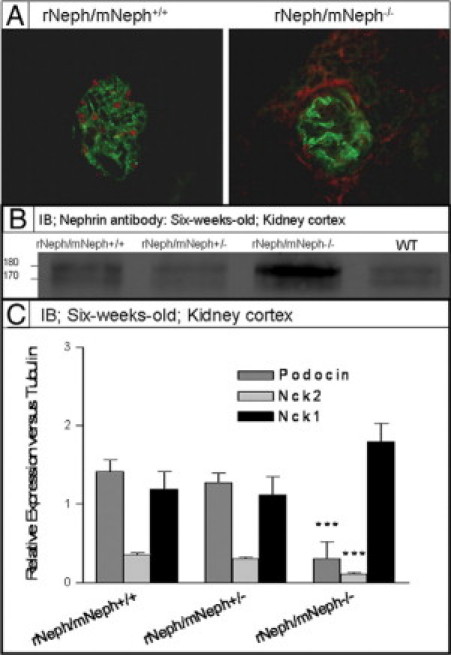
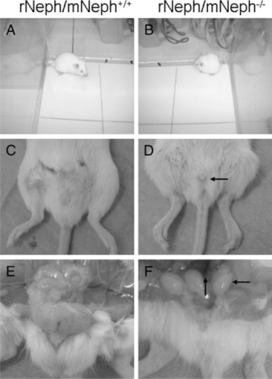
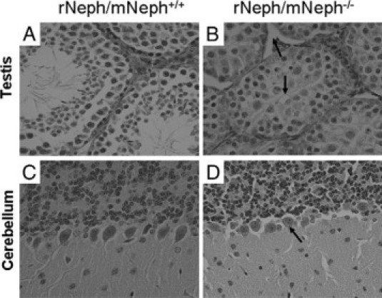
Mutations leading to nephrin loss result in massive proteinuria both in humans and mice. Early perinatal lethality of conventional nephrin knockout mice makes it impossible to determine the role of nephrin protein in the adult kidney and in extra-renal tissues. Herein, we studied whether podocyte-specific, doxycycline-inducible, rat nephrin expression can rescue nephrin-deficient mice from perinatal lethality. Fourteen littermates out of 72 lacked endogenous nephrin and expressed transgenic rat nephrin. Six of these rescued mice survived until 6 weeks of age, whereas the nephrin-deficient pups died before the age of 5 days. The rescued mice were smaller, developed proteinuria, and showed histological abnormalities in the kidney. Despite foot process effacement, slit diaphragms were observed. Importantly, the expression and localization of several proteins associated with the signaling capacity of nephrin or the regulation of the expression of nephrin were changed in the podocytes. Indeed, all rescued mice showed impaired locomotor activity and distinct histological abnormalities in the cerebellum, and the male mice were also infertile and showed genital malformations. These observations are consistent with normal nephrin expression in the testis and cerebellum. These observations indicate that podocyte-specific expression of rat nephrin can rescue nephrin-deficient mice from perinatal death, but is not sufficient for full complementation.


Figures

Figure 1
Figure 1
The rNephrin transgenic mouse. A: The JRC-rNephrin transgene construct: In a binary reverse doxycycline-regulated expression vector an NPHS2 (podocin) promoter drives the expression of the recombinant inducible transcription factor reverse tetracycline transactivator (M2). In the presence of doxycycline, reverse tetracycline transactivator binds to the tetO7 element linked to a minimal CMV promoter that drives the expression of rat nephrin cDNA. B: Rat- and mouse-specific nephrin mRNA expressions in JRC-rNephrin (rNeph) mouse kidneys isolated from nine founder-lines (93A – 66A) analyzed by RT-PCR. C: RT-PCR of mRNA from the kidney cortex of the selected JRC-rNephrin founder-line (86A): 1) Wild-type mouse without doxycycline, 2) rNeph mouse without doxycycline, and 3) rNeph mouse with doxycycline. Total RNA was isolated from kidneys collected from adult mice at the age of 10 to 12 weeks after 2 weeks of doxycycline (0.2 mg/ml) administration. PCR was performed using primers presented in Table 2. Similar results were obtained with three independent experiments with three mice in each genotype.
Figure 2
Figure 2
Expressions of rat nephrin, mouse nephrin, podocin, and WT1 mRNAs analyzed by RT-PCR. Total RNA was isolated from the kidneys of E11 and E14 wild-type (WT) and rNeph embryos collected from pregnant rNeph transgenic mouse line females with 2.0 mg/ml doxycycline in the drinking water. The primers used are listed in Table 2. Similar results were obtained with three independent experiments with three mice in each genotype.
Figure 3
Figure 3
Expression of transgenic rat nephrin in podocytes rescues conventional nephrin KO mice from perinatal lethality with normal kidney and podocyte morphology at the age of 1 week. A: Mouse- and rat-specific nephrin mRNA expressions in rNeph/mNeph mice kidney cortex at the age of 1 week, as analyzed by RT-PCR. Genotypes are marked 1) rNeph/mNeph+/+, (2) rNeph/mNeph−/− and (3) rNeph/mNeph+/−. B and C: Normal nephrin protein expression and localization in rNeph/mNeph+/+ and rNeph/mNeph−/− mice at the age of 1 week analyzed by immunofluorescent microscopy of frozen kidney sections (magnification ×100). D and E: Kidney histology in rNeph/mNeph+/+ and rNeph/mNeph−/− mice at the age of 1 week analyzed by H&E staining of paraffin-embedded kidney sections (×200 magnification). F and G: Electron microscopy shows relatively normal podocyte fine structures and well-preserved slit diaphragm structures in kidneys of rNeph/mNeph+/+ and rNeph/mNeph−/− mice. Arrows indicate existing slit diaphragms (SDs). Glomerular basement membrane (GBM); urinary space (US). Scale bars = 200 nm.
Figure 4
Figure 4
The kidneys from 6-week-old rescued rNeph/mNeph−/− mice showed proteinuria-associated histological abnormalities and a variety of podocyte damage. A and B: H&E staining of paraffin-embedded kidney sections from rNeph/mNeph+/+ and rNeph/mNeph−/− mice (magnification ×200). C–F: Electron microscopy showed a variety of morphological changes such as a foot process effacement, mesangial expansion, and glomerular basement membrane thickening in podocytes of the rescued rNeph/mNeph−/− mice, as compared with rNeph/mNeph+/+ littermate controls. Despite of foot process effacement, slit diaphragm structures were still observed in rNeph/mNeph−/− mice (F). Arrows indicate still existing slit diaphragms (SDs). Asterisks indicate area were partial foot process effacement can be seen. Glomerular basement membrane (GBM), urinary space (US). Scale bars: 5 μm (C and D); 200 nm (E and F).
Figure 5
Figure 5
The expression and localization of the rat nephrin mRNA and protein in the kidneys was similar in rescued rNeph/mNeph−/− mice, as compared with control littermates at the age of 6 weeks. A: Mouse- and rat-specific nephrin mRNA expressions in rNeph/mNeph mice kidney cortex at the age of 6 weeks, as analyzed by RT-PCR. Genotypes are marked 1) rNeph/mNeph+/+, (2) rNeph/mNeph−/−, and (3) rNeph/mNeph+/−. B and C: Nephrin protein expression and localization in rNeph/mNeph+/+ and rNeph/mNeph−/− mice at the age of 6 weeks, analyzed by immunofluorescent microscopy of frozen kidney sections (magnification ×200). D and E: Immunogold electron microscopy shows normal nephrin localization in the slit diaphragm area in rescued rNeph/mNeph−/− mice, when compared with control rNeph/mNeph+/+ mice. Arrows indicate nephrin protein localization. Glomerular basement membrane (GBM). F: Nephrin localization was unaltered in slit diaphragm area and in podocyte cytoplasm in the rescued rNeph/mNeph−/− mouse, as compared with the control rNeph/mNeph+/+ mouse. However, more nephrin protein was found in the podocyte membrane outside the slit diaphragm area from the rescued mouse and this increase was statistically significant (*P < 0.05). The number of gold particles were counted manually and expressed as the number/1000 nm length of podocyte membrane. Student's t-test was used for statistical analysis.
Figure 6
Figure 6
Changes expression of podocin, Nck2, and WT1 mRNAs in 6-week-old rescued rNeph/mNeph−/− mice. RT-PCR was performed with total RNA isolated from kidney cortex at the age of 6 weeks. Genotypes are marked: 1) rNeph/mNeph+/+, 2) rNeph/mNeph−/−, and 3) rNeph/mNeph+/−. A and B: Podocin and Nck 2 expression was decreased in rescued rNeph/mNeph−/− mice kidneys. C: WT1 expression was analyzed using a primer set designed to amplify different splicing forms with or without exon 5. The results showed an increase in the exon 5 containing mRNA variant in rescued rNeph/mNeph−/− mice, as compared with the control genotypes. D: β-actin expression was similar in all genotypes. Primers and size of the products are listed in Table 2. Similar results were obtained in three independent experiments with three mice of each genotype.
Figure 7
Figure 7
Changed podocin and Fyn protein expression in 6-week-old rescued rNeph/mNeph−/− mice. Immunofluorescent double-staining of frozen kidney sections from rNeph/mNeph+/+ (left panel) and rNeph/mNeph−/− (right panel) mice with podocin-, Fyn-, and synaptopodin-specific antibodies showed that podocin protein is markedly decreased in the rescued mice, as compared with control mouse kidneys (two upper rows). Instead, the Fyn protein levels in the area close to the podocyte foot processes and slit diaphragm were increased in the rescued mice (two lowest rows). Magnification: ×200.
Figure 8
Figure 8
Absence of Nck2 protein expression in 6-week-old rescued rNeph/mNeph−/− mice. Immunofluorescent staining of frozen kidney sections from rNeph/mNeph+/+ (left panel) and rNeph/mNeph−/− (right panel) mouse kidneys with Nck1-, Nck2-, and synaptopodin-specific antibodies showed slit diaphragm-specific staining and co-localization of Nck2 protein with synaptopodin in rNeph/mNeph+/+ mice (two lower rows). The Nck1 protein shows some glomerular, but also strong tubular staining (two upper rows). The Nck1 protein expression levels remain closely similar between genotypes, but Nck2 protein was completely absent in rNeph/mNeph−/− mice. Magnification: ×200.
Figure 9
Figure 9
A: Immunofluorescent staining with WT1 and synaptopodin specific antibodies on frozen kidney sections from 6-week-old rNeph/mNeph+/+, and rescued rNeph/mNeph−/− mice. Surprisingly, WT1 expression was absent in the nucleus of 6-week-old rescued rNeph/mNeph−/− mice podocytes when still present in age-matched control kidneys. Magnification: ×200. B: Immunoblotting from kidney cortex lysate with nephrin antibody showed an increased level of the phosphorylated nephrin form (∼180 kDa) rNeph/mNeph−/− mice, as compared with the other genotypes (Figure 9B). C: Quantitative immunoblotting experiments verified RT-PCR and immunofluorescent microscopic findings that expression level of podocin and Nck2 has been decreased (***P < 0.001) and Nck1 unchanged in rescued mice as compared with control rNeph/mNeph+/+ littermates.
Figure 10
Figure 10
All rescued rNeph/mNeph−/− male mice were smaller in size, infertile, and their genitals showed anatomical malformations at the age of 5 to 6 weeks. A and B show the differences in body size. C and D show the smaller size of the male genitals of rNeph/mNeph−/− mice. E and F show that testicles had not descended and seminal vesicle formation was severely impaired in rNeph/mNeph−/− males. These changes have been highlighted with arrows in D and F.
Figure 11
Figure 11
Histological experiments by H&E staining and light microscopy revealed striking morphological differences in testis and cerebellum of all rescued rNeph/mNeph−/− male mice at the age of 6 weeks. These histological changes included (marked as arrows): A and B: Decreased number of Leydig cells and spermatids and absence of the lumen of the seminiferous tubules of the testis. C and D: The loss of polarization of the Purkinje cells in the cerebellum. Magnification: ×200.

Similar articles

Cited by

References

    1. Rapola J. Congenital nephrotic syndrome. Pediatr Nephrol. 1987;1:441–446. - PubMed
    1. Kestila M, Lenkkeri U, Mannikko M, Lamerdin J, McCready P, Putaala H, Ruotsalainen V, Morita T, Nissinen M, Herva R, Kashtan CE, Peltonen L, Holmberg C, Olsen A, Tryggvason K. Positionally cloned gene for a novel glomerular protein–nephrin–is mutated in congenital nephrotic syndrome. Mol Cell. 1998;1:575–582. - PubMed
    1. Lenkkeri U, Mannikko M, McCready P, Lamerdin J, Gribouval O, Niaudet PM, Antignac CK, Kashtan CE, Homberg C, Olsen A, Kestila M, Tryggvason K. Structure of the gene for congenital nephrotic syndrome of the finnish type (NPHS1) and characterization of mutations. Am J Hum Genet. 1999;64:51–61. - PMC - PubMed
    1. Hallman N, Hjelt L. Congenital nephrotic syndrome. J Pediatr. 1959;55:152–162. - PubMed
    1. Tryggvason K, Kouvalainen K. Number of nephrons in normal human kidneys and kidneys of patients with the congenital nephrotic syndrome. A study using a sieving method for counting of glomeruli. Nephron. 1975;15:62–68. - PubMed

Publication types

MeSH terms