

Endothelial progenitor cells (EPCs) mobilized and activated by neurotrophic factors may contribute to pathologic neovascularization in diabetic retinopathy
- PMID: 19948824
- PMCID: PMC2797908
- DOI: 10.2353/ajpath.2010.081152
Endothelial progenitor cells (EPCs) mobilized and activated by neurotrophic factors may contribute to pathologic neovascularization in diabetic retinopathy
Abstract
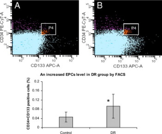
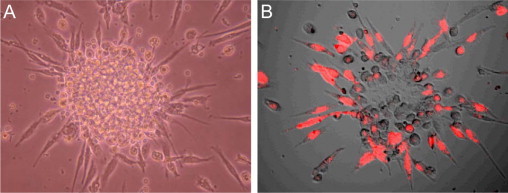
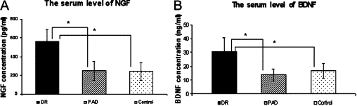
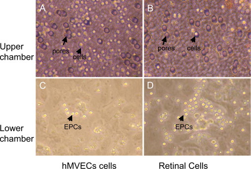
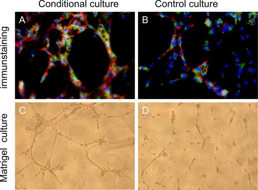
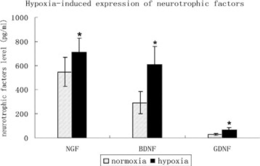
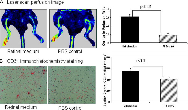
Diabetic retinopathy is characterized by pathological retinal neovascularization. Accumulating evidence has indicated that high levels of circulating endothelial progenitor cells (EPCs) are an important risk factor for neovascularization. Paradoxically, the reduction and dysfunction of circulating EPCs has been extensively reported in diabetic patients. We hypothesized that EPCs are differentially altered in the various vasculopathic complications of diabetes mellitus, exhibiting distinct behaviors in terms of angiogenic response to ischemia and growth factors and potentially playing a potent role in motivating vascular precursors to induce pathological neovascularization. Circulating levels of EPCs from diabetic retinopathy patients were analyzed by flow cytometry and by counting EPC colony-forming units, and serum levels of neurotrophic factors were measured by enzyme-linked immunosorbent assay. We found increased levels of nerve growth factor and brain-derived neurotrophic factor in the blood of diabetic retinopathy patients; this increase was correlated with the levels of circulating EPCs. In addition, we demonstrated that retinal cells released neurotrophic factors under hypoxic conditions to enhance EPC activity in vitro and to increase angiogenesis in a mouse ischemic hindlimb model. These results suggest that neurotrophic factors may induce neoangiogenesis through EPC activation, leading to the pathological retinal neovascularization. Thus, we propose that neovascularization in the ischemic retina might be regulated by overexpression of neurotrophic factors.
Figures
Similar articles
-
Endothelial precursor cells promote angiogenesis in hepatocellular carcinoma.World J Gastroenterol. 2012 Sep 21;18(35):4925-33. doi: 10.3748/wjg.v18.i35.4925. World J Gastroenterol. 2012. PMID: 23002366 Free PMC article.
-
The involvement of endothelial progenitor cells in tumor angiogenesis.J Cell Mol Med. 2004 Jul-Sep;8(3):294-300. doi: 10.1111/j.1582-4934.2004.tb00319.x. J Cell Mol Med. 2004. PMID: 15491505 Free PMC article. Review.
-
CD34 hybrid cells promote endothelial colony-forming cell bioactivity and therapeutic potential for ischemic diseases.Arterioscler Thromb Vasc Biol. 2013 Jul;33(7):1622-34. doi: 10.1161/ATVBAHA.112.301052. Epub 2013 May 2. Arterioscler Thromb Vasc Biol. 2013. PMID: 23640491
-
Circulating endothelial progenitor cells in systemic sclerosis: relation to impaired angiogenesis and cardiovascular manifestations.Clin Exp Rheumatol. 2008 May-Jun;26(3):421-9. Clin Exp Rheumatol. 2008. PMID: 18578963
-
Endothelial progenitor cells in angiogenesis.Sheng Li Xue Bao. 2005 Feb 25;57(1):1-6. Sheng Li Xue Bao. 2005. PMID: 15719128 Review.
Cited by
-
Potential role of intravitreal human placental stem cell implants in inhibiting progression of diabetic retinopathy in type 2 diabetes: neuroprotective growth factors in the vitreous.Clin Ophthalmol. 2011;5:691-6. doi: 10.2147/OPTH.S21161. Epub 2011 May 23. Clin Ophthalmol. 2011. PMID: 21629576 Free PMC article.
-
Comparison of three fluorescence labeling and tracking methods of endothelial progenitor cells in laser-injured retina.Int J Ophthalmol. 2018 Apr 18;11(4):580-588. doi: 10.18240/ijo.2018.04.07. eCollection 2018. Int J Ophthalmol. 2018. PMID: 29675374 Free PMC article.
-
Nerve Growth Factor Promotes Angiogenesis and Skeletal Muscle Fiber Remodeling in a Murine Model of Hindlimb Ischemia.Chin Med J (Engl). 2016 Feb 5;129(3):313-9. doi: 10.4103/0366-6999.174496. Chin Med J (Engl). 2016. PMID: 26831234 Free PMC article.
-
Stem cell therapy for abrogating stroke-induced neuroinflammation and relevant secondary cell death mechanisms.Prog Neurobiol. 2017 Nov;158:94-131. doi: 10.1016/j.pneurobio.2017.07.004. Epub 2017 Jul 23. Prog Neurobiol. 2017. PMID: 28743464 Free PMC article. Review.
-
Leverage of nuclease-deficient CasX for preventing pathological angiogenesis.Mol Ther Nucleic Acids. 2023 Aug 6;33:738-748. doi: 10.1016/j.omtn.2023.08.001. eCollection 2023 Sep 12. Mol Ther Nucleic Acids. 2023. PMID: 37662968 Free PMC article.
References
-
- Asahara T, Murohara T, Sullivan A, Silver M, van der Zee R, Li T, Witzenbichler B, Schatteman G, Isner JM. Isolation of putative progenitor endothelial cells for angiogenesis. Science. 1997;275:964–967. - PubMed
-
- Takahashi T, Kalka C, Masuda H, Chen D, Silver M, Kearney M, Magner M, Isner JM, Asahara T. Ischemia- and cytokine-induced mobilization of bone marrow-derived endothelial progenitor cells for neovascularization. Nat Med. 1999;5:434–438. - PubMed
-
- Otani A, Kinder K, Ewalt K, Otero FJ, Schimmel P, Friedlander M. Bone marrow-derived stem cells target retinal astrocytes and can promote or inhibit retinal angiogenesis. Nat Med. 2002;8:1004–1010. - PubMed
-
- Grant MB, May WS, Caballero S, Brown GA, Guthrie SM, Mames RN, Byrne BJ, Vaught T, Spoerri PE, Peck AB, Scott EW. Adult hematopoietic stem cells provide functional hemangioblast activity during retinal neovascularization. Nat Med. 2002;8:607–612. - PubMed
-
- Lee IG, Chae SL, Kim JC. Involvement of circulating endothelial progenitor cells and vasculogenic factors in the pathogenesis of diabetic retinopathy. Eye. 2006;20:546–552. - PubMed
Publication types
MeSH terms
Substances
LinkOut - more resources
Full Text Sources
Medical
