

The role of liver sinusoidal cells in hepatocyte-directed gene transfer
- PMID: 19948827
- PMCID: PMC2797863
- DOI: 10.2353/ajpath.2010.090136
The role of liver sinusoidal cells in hepatocyte-directed gene transfer
Abstract
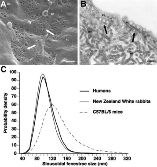
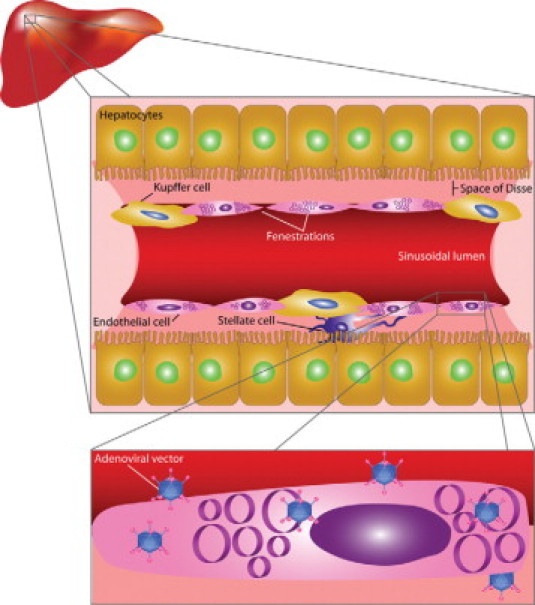
Hepatocytes are a key target for gene therapy of inborn errors of metabolism as well as of acquired diseases such as liver cancer and hepatitis. Gene transfer efficiency into hepatocytes is significantly determined by histological and functional aspects of liver sinusoidal cells. On the one hand, uptake of vectors by Kupffer cells and liver sinusoidal endothelial cells may limit hepatocyte transduction. On the other hand, the presence of fenestrae in liver sinusoidal endothelial cells provides direct access to the space of Disse and allows vectors to bind to receptors on the microvillous surface of hepatocytes. Nevertheless, the diameter of fenestrae may restrict the passage of vectors according to their size. On the basis of lege artis measurements of the diameter of fenestrae in different species, we show that the diameter of fenestrae affects the distribution of transgene DNA between sinusoidal and parenchymal liver cells after adenoviral transfer. The small diameter of fenestrae in humans may underlie low efficiency of adenoviral transfer into hepatocytes in men. The disappearance of the unique morphological features of liver sinusoidal endothelial cells in pathological conditions like liver cirrhosis and liver cancer may further affect gene transfer efficiency. Preclinical gene transfer studies should consider species differences in the structure and function of liver sinusoidal cells as important determinants of gene transfer efficiency into hepatocytes.
Figures
References
-
- Do H, Healey JF, Waller EK, Lollar P. Expression of factor VIII by murine liver sinusoidal endothelial cells. J Biol Chem. 1999;274:19587–19592. - PubMed
-
- Shiratori Y, Tananka M, Kawase T, Shiina S, Komatsu Y, Omata M. Quantification of sinusoidal cell function in vivo. Semin Liver Dis. 1993;13:39–49. - PubMed
-
- Bergelson JM, Cunningham JA, Droguett G, Kurt-Jones EA, Krithivas A, Hong JS, Horwitz MS, Crowell RL, Finberg RW. Isolation of a common receptor for Coxsackie B viruses and adenoviruses 2 and 5. Science. 1997;275:1320–1323. - PubMed
Publication types
MeSH terms
LinkOut - more resources
Full Text Sources
Other Literature Sources
