. 2010 Jan;176(1):218-26.
doi: 10.2353/ajpath.2010.081027. Epub 2009 Nov 30.

CD11c+ cells are required to prevent progression from local acute lung injury to multiple organ failure and death

Affiliations

CD11c+ cells are required to prevent progression from local acute lung injury to multiple organ failure and death

Jami E Milam et al. Am J Pathol. 2010 Jan.

Abstract

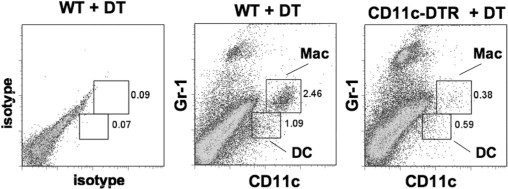
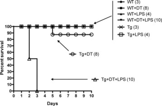
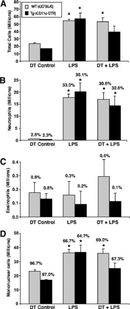
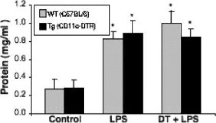
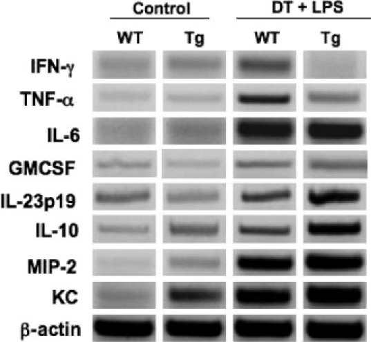
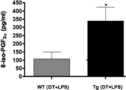
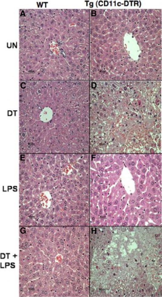
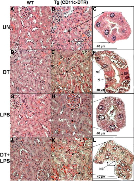
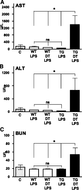
To investigate the role of CD11c(+) cells in endotoxin-induced acute lung injury, wild-type or CD11c-diphtheria toxin receptor transgenic mice were treated with intraperitoneal diphtheria toxin (5 ng/g b.wt.) in the presence or absence of intratracheal lipopolysaccharide (51 microg). Lipopolysaccharide treatment resulted in 100% mortality in CD11c-depleted animals but not in control animals. Analysis of local lung tissue revealed no differences in acute lung injury severity; however, analysis of distal tissues revealed severe damage and necrosis to multiple organs (liver, spleen, and kidneys) in CD11c-diphtheria toxin receptor mice but not in wild-type mice. In addition, dramatic increases in systemic levels of liver enzymes (alanine aminotransferase, 657 U/L, aspartate aminotransferase, 1401 U/L), blood urea (53 mg/dl), and 8-iso-prostaglandin F(2alpha), a marker of oxidative stress (350 pg/ml), were observed. These data demonstrate that CD11c(+) cells play a critical role in protecting the organs from systemic injury caused by a pulmonary endotoxin challenge.


Figures

Figure 1
Figure 1
Effect of DT on CD11c depletion in the lungs of wild-type (WT) and CD11c-DTR mice. This representative graph is a gray-scale pseudo-color plot of lung leukocytes from wild-type and C57BL/6 CD11c-DTR mice harvested 24 hours after DT treatment and then stained with control antibodies (left panel), or antibodies against CD11c and Gr-1 (middle and right panels).
Figure 2
Figure 2
Effect of DT and LPS on survival in C57BL/6 wild-type (WT) versus CD11c-DTR Tg mice of the same background. The mice were treated i.p. with 5 ng/g b.wt. DT on day −1 and/or intratracheally inoculated with (51 μg) of LPS on day 0. The number in parentheses indicates the number of animals/treatment group.
Figure 3
Figure 3
Comparison of lung leukocyte recruitment in response to LPS challenge in wild-type (WT) C57BL/6 and Tg CD11c-DTR mice. Samples of individual lung leukocyte suspensions were spun onto slides and stained with Wright-Giemsa stain for visual quantification of leukocyte subsets as described in Materials and Methods. A: Total cells. B: Neutrophils. C: Eosinophils. D: Mononuclear cells. *P < 0.05 compared with the untreated mice. n = 3 to 11 mice/group.
Figure 4
Figure 4
The comparison of severity in alveolar-capillary leak as reflected by the protein concentration of bronchoalveolar lavage fluid in wild-type (WT) C57BL/6 and Tg CD11c-DTR mice with or without DT and LPS treatment compared with LPS alone. *P < 0.05 compared with the untreated mice of the same group. n = 3 to 12 mice/group; the specific number of animals per group is described in Materials and Methods.
Figure 5
Figure 5
RT-PCR analysis of interferon-γ (IFN-γ), tumor necrosis factor-α (TNFα), IL-10, IL-6, IL-17, IL-23p19, granulocyte-macrophage colony-stimulating factor (GMCSF), MIP-2, KC, and β-actin in the whole lung homogenates from wild-type (WT) C57BL/6 and Tg CD11c-DTR mice of the same background. Each lane is a representative of at least three animals per group.
Figure 6
Figure 6
Markers of oxidative stress/injury in wild-type (WT) and BALB/c CD11c-DTR mice. The total 8-isoprostane-prostagandin F (8-iso-PGF) level, a marker of oxidative stress and injury, was measured on day 2 after LPS administration in the fresh plasma of mice treated with DT + LPS. *P < 0.05. n = 5 mice.
Figure 7
Figure 7
Photomicrographs of liver sections showing necrosis in LPS- and DT + LPS-treated Tg (CD11c-DTR) mice that were harvested on day 1. Wild-type BALB/c mice (A, C, E, and G), labeled (WT), and the Tg (CD11c-DTR) mice of the same background (B, D, F, and H), labeled Tg (CD11c-DTR): untreated (A and B), labeled (UN); or treated i.p. with 5 ng/g b.wt. with DT (C and D) day −1, labeled (DT); or intratracheally inoculated with (51 μg) of LPS (E and F) day 0, labeled (LPS); or treated with both DT and LPS (G and H) and harvested on day 1, labeled (DT + LPS). The liver sections were stained with H&E and examined under a microscope (×40). Livers from the Tg (CD11c-DTR) mice treated with DT (D) show liver necrosis including fatty/vacuolar dystrophy and red blood cell infiltration. Livers from the Tg (CD11c-DTR) mice treated with DT + LPS (H) show extensive liver necrosis including fatty/vacuolar dystrophy, red blood cell infiltration, and hepatocyte disintegration.
Figure 8
Figure 8
Photomicrographs of kidney sections showing necrosis in DT- and DT + LPS-treated Tg (CD11c-DTR) mice that were harvested on day 1. The wild-type BALB/c mice (A, D, G, and J), labeled (WT), and the Tg (CD11c-DTR) mice of the same background (B, E, H, and K), labeled Tg (CD11c-DTR), and also a enlarged multicellular tubule unit (C, F, I, and L): untreated (A, B, and C), labeled (UN); or treated i.p. with 5 ng/g b.wt. with DT (D, E, and F) day −1, labeled (DT); or intratracheally inoculated with (51 μg) of LPS (G, H, and I) on day 0, labeled (LPS); or treated with both DT and LPS (J, K, and L) and harvested on day 1, labeled (DT + LPS). The kidney sections were stained with H&E and examined under a microscope (×40). The arrows with dots on the ends show where the enlarged groups of cells were located. Kidney from the Tg (CD11c-DTR) mice treated with DT (E and F) shows necrosis (NE) of the tubules with some normal (N) cells. Kidney from the Tg (CD11c-DTR) mice treated with DT + LPS (K and L) shows extensive necrosis (NE) of the tubules and renal cell disintegration. Tg (CD11c-DTR) figure sets with DT (E and F) and DT + LPS (K and L) have a brown color compared with the normal pink/purple. Scale bar = 40 μm.
Figure 9
Figure 9
Activity of circulating alanine aminotransferase (ALT) (A) and aspartate aminotransferase (AST) (B) and blood urea nitrogen (BUN) (C) in LPS- and DT + LPS-treated BALB/c CD11c-DTR and wild-type (WT) mice. Blood was collected via orbital bleeding on day 2 after LPS challenge. Data as means ± SE. *P < 0.05.

Similar articles

Cited by

References

    1. Ware LB, Matthay MA. The acute respiratory distress syndrome. N Engl J Med. 2000;342:1334–1349. - PubMed
    1. Ware LB. Pathophysiology of acute lung injury and the acute respiratory distress syndrome. Semin Respir Crit Care Med. 2006;27:337–349. - PubMed
    1. Reutershan J, Ley K. Bench-to-bedside review: acute respiratory distress syndrome-how neutrophils migrate into the lung. Crit Care. 2004;8:453–461. - PMC - PubMed
    1. Frutos-Vivar F, Ferguson ND, Esteban A. Epidemiology of acute lung injury and acute respiratory distress syndrome. Semin Respir Crit Care Med. 2006;27:327–336. - PubMed
    1. Bhatia M, Moochhala S. Role of inflammatory mediators in the pathophysiology of acute respiratory distress syndrome. J Pathol. 2004;202:145–156. - PubMed

Publication types

MeSH terms