. 2010 Feb;78(2):704-15.
doi: 10.1128/IAI.00881-09. Epub 2009 Nov 30.

The Streptococcus pneumoniae capsule inhibits complement activity and neutrophil phagocytosis by multiple mechanisms

Affiliations

The Streptococcus pneumoniae capsule inhibits complement activity and neutrophil phagocytosis by multiple mechanisms

Catherine Hyams et al. Infect Immun. 2010 Feb.

Abstract

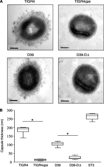
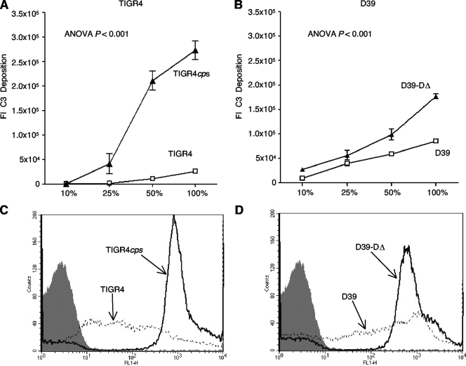
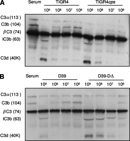
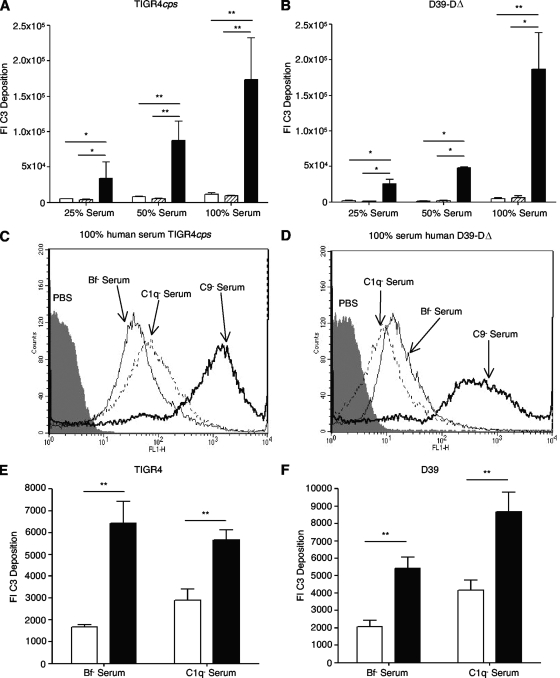
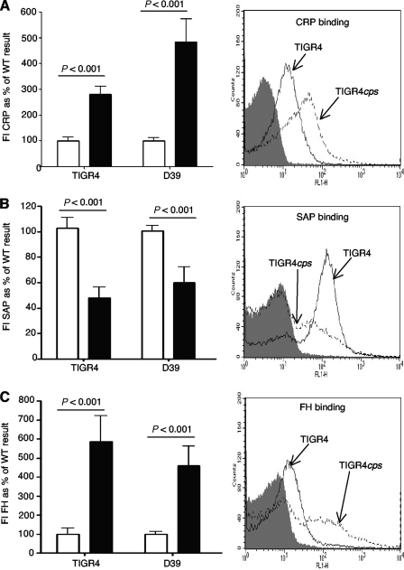
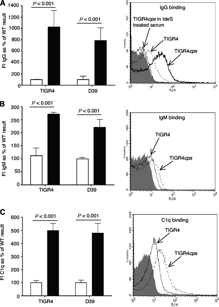
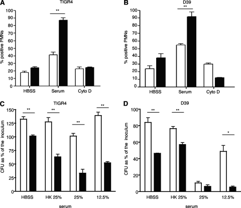
The Streptococcus pneumoniae capsule is vital for virulence and may inhibit complement activity and phagocytosis. However, there are only limited data on the mechanisms by which the capsule affects complement and the consequences for S. pneumoniae interactions with phagocytes. Using unencapsulated serotype 2 and 4 S. pneumoniae mutants, we have confirmed that the capsule has several effects on complement activity. The capsule impaired bacterial opsonization with C3b/iC3b by both the alternative and classical complement pathways and also inhibited conversion of C3b bound to the bacterial surface to iC3b. There was increased binding of the classical pathway mediators immunoglobulin G (IgG) and C-reactive protein (CRP) to unencapsulated S. pneumoniae, indicating that the capsule could inhibit classical pathway complement activity by masking antibody recognition of subcapsular antigens, as well as by inhibiting CRP binding. Cleavage of serum IgG by the enzyme IdeS reduced C3b/iC3b deposition on all of the strains, but there were still marked increases in C3b/iC3b deposition on unencapsulated TIGR4 and D39 strains compared to encapsulated strains, suggesting that the capsule inhibits both IgG-mediated and IgG-independent complement activity against S. pneumoniae. Unencapsulated strains were more susceptible to neutrophil phagocytosis after incubation in normal serum, normal serum treated with IdeS, complement-deficient serum, and complement-deficient serum treated with IdeS or in buffer alone, suggesting that the capsule inhibits phagocytosis mediated by Fcgamma receptors, complement receptors, and nonopsonic receptors. Overall, these data show that the S. pneumoniae capsule affects multiple aspects of complement- and neutrophil-mediated immunity, resulting in a profound inhibition of opsonophagocytosis.


Figures

FIG. 1.
FIG. 1.
Measurement of capsule layer diameter by EM. (A) Examples of EM of encapsulated and unencapsulated strains. Bars, 200 nm. (B) Capsule layer widths (nm) for the TIGR4 and D39 strains, their unencapsulated counterparts TIGR4cps and D39-DΔ, and a capsular serotype 3 strain, presented as medians and interquartile ranges. For comparisons between unencapsulated and encapsulated strains, an asterisk indicates a P value of <0.001 (Mann-Whitney U test).
FIG. 2.
FIG. 2.
Effect of the S. pneumoniae capsule on C3b/iC3b deposition. (A and B) FI of C3b/iC3b deposition measured using a flow cytometry assay on the TIGR4 (A) and D39 (B) strains for encapsulated (□) and unencapsulated strains (TIGR4cps and D39-DΔ) (▴) in increasing concentrations of human serum. (C) and (D) Examples of flow cytometry histograms for C3b/iC3b deposition on TIGR4 and D39 wild-type and unencapsulated strains in 100% human serum. Gray shading indicates the results for bacteria incubated in PBS alone. In panels A and B, error bars represent SDs, and for the differences between encapsulated and unencapsulated organisms, the Ρ value is <0.001 (ANOVA).
FIG. 3.
FIG. 3.
Representative immunoblot assays of serum incubated with differing numbers of CFU of encapsulated and unencapsulated TIGR4 (A) and D39 (B) bacteria and then probed with an Ab to C3 and its breakdown products. The positions of breakdown products of C3 (with their approximate sizes in kDa in parentheses) are shown to the left of each panel. Similar results were obtained with repeated immunoblot assays.
FIG. 4.
FIG. 4.
Effects of classical and alternative pathways on C3b/iC3b deposition on unencapsulated strains. (A and B) FI for flow cytometry results of C3b/iC3b deposition on the TIGR4cps (A) and D39-DΔ (B) strains in different concentrations of human serum depleted of C9 (black bars), C1q (open bars), or Bf (diagonally shaded bars). (C and D) Examples of flow cytometry histograms for C3b/iC3b deposition on strain TIGR4cps (C) and D39-DΔ (D) bacteria incubated in 100% C9 (thick black line), C1q (thin black line), or Bf (dashed line) human serum. (E) Comparison of the FIs for C3b/iC3b deposition in C1q (C1q)- or Bf (Bf)-depleted serum on the TIGR4 (white bars) and TIGR4cps (black bars) strains. (F) Comparison of the FIs for C3b/iC3b deposition in C1q (C1q)- or Bf (Bf)-depleted serum on the D39 (white bars) and D39-DΔ (black bars) strains. In panels A, B, E, and F, error bars represent SDs and single and double asterisks indicate a P value of < 0.01 and < 0.001, respectively (ANOVAs with post-hoc analysis).
FIG. 5.
FIG. 5.
Flow cytometry assays of binding of the pentraxins CRP and SAP and the alternative pathway inhibitor FH to unencapsulated and encapsulated S. pneumoniae in 50% human serum. (A) FIs of CRP binding to the TIGR4 and D39 strains (white bars), compared to the TIGR4cps and D39-DΔ strains (black bars), and an example of the flow cytometry histogram for the TIGR4 strains. (B) FIs of SAP binding to the TIGR4 and D39 strains (white bars), compared to the TIGR4cps and D39-DΔ strains (black bars), and an example of the flow cytometry histogram for the TIGR4 strains. (C) FIs of FH binding to the TIGR4 and D39 strains (white bars), compared to the TIGR4cps and D39-DΔ strains (black bars), and an example of the flow cytometry histogram for the TIGR4 strains. In all panels, error bars represent SDs and P values were obtained using unpaired Student t tests. For the representative flow cytometry histograms, gray shading indicates the results for TIGR4cps incubated in PBS alone.
FIG. 6.
FIG. 6.
Binding of IgG, IgM, and C1q to unencapsulated S. pneumoniae in 50% human serum. (A) FIs of IgG binding to the TIGR4 and D39 strains (white bars), compared to the TIGR4cps and D39-DΔ strains (black bars) and an example of the flow cytometry histogram for the TIGR4 strains, including an example of the results obtained with serum treated with IdeS. (B) FIs of IgM binding to the TIGR4 and D39 strains (white bars), compared to the TIGR4cps and D39-DΔ strains (black bars), and an example of the flow cytometry histogram for the TIGR4 strains. (C) FIs of C1q binding to the TIGR4 and D39 strains (white bars), compared to the TIGR4cps and D39-DΔ strains (black bars), and an example of the flow cytometry histogram for the TIGR4 strains. In all panels, error bars represent SDs and P values were obtained using Student unpaired t tests. WT, wild type.
FIG. 7.
FIG. 7.
Effect of the capsule on the interactions of S. pneumoniae with human neutrophils. (A and B) Percent association of fresh human neutrophils with strain TIGR4 (A) or D39 (B) (open bars) and strain TIGR4cps or D39-DΔ (black bars) when opsonized in 20% serum with or without inhibition of phagocytosis using cytochalasin D. Error bars represent SDs, and single and double asterisks represent Ρ values of < 0.01 and < 0.001, respectively (ANOVA with post-hoc tests). (C and D) Proportion of S. pneumoniae inoculum surviving after incubation with fresh human neutrophils for 30 min when opsonized with 12.5 or 25% human serum, 25% heat-treated serum (HK), or HBSS. (C) Results for strains TIGR4 (white bars) and TIGR4cps (black bars). (D) Results for strains D39 (white bars) and D39-DΔ (black bars). Error bars represent SDs, and single and double asterisks represent Ρ values of < 0.05 and < 0.01, respectively, for comparisons of unencapsulated to encapsulated bacteria under each opsonization condition (Student t test). PMN, polymorphonuclear neutrophils; WT, wild type.

Similar articles

Cited by

References

    1. Abeyta, M., G. G. Hardy, and J. Yother. 2003. Genetic alteration of capsule type but not PspA type affects accessibility of surface-bound complement and surface antigens of Streptococcus pneumoniae. Infect. Immun. 71:218-225. - PMC - PubMed
    1. Arredouani, M., Z. Yang, Y. Ning, G. Qin, R. Soininen, K. Tryggvason, and L. Kobzik. 2004. The scavenger receptor MARCO is required for lung defense against pneumococcal pneumonia and inhaled particles. J. Exp. Med. 200:267-272. - PMC - PubMed
    1. Arredouani, M. S., Z. Yang, A. Imrich, Y. Ning, G. Qin, and L. Kobzik. 2006. The macrophage scavenger receptor SR-AI/II and lung defense against pneumococci and particles. Am. J. Respir. Cell Mol. Biol. 35:474-478. - PMC - PubMed
    1. Baxendale, H. E., M. Johnson, R. C. Stephens, J. Yuste, N. Klein, J. S. Brown, and D. Goldblatt. 2008. Natural human antibodies to pneumococcus have distinctive molecular characteristics and protect against pneumococcal disease. Clin. Exp. Immunol. 151:51-60. - PMC - PubMed
    1. Bentley, S. D., D. M. Aanensen, A. Mavroidi, D. Saunders, E. Rabbinowitsch, M. Collins, K. Donohoe, D. Harris, L. Murphy, M. A. Quail, G. Samuel, I. C. Skovsted, M. S. Kaltoft, B. Barrell, P. R. Reeves, J. Parkhill, and B. G. Spratt. 2006. Genetic analysis of the capsular biosynthetic locus from all 90 pneumococcal serotypes. PLoS Genet. 2:e31. - PMC - PubMed

Publication types

MeSH terms

Substances