. 2010 Feb;78(2):716-25.
doi: 10.1128/IAI.01056-09. Epub 2009 Nov 30.

Streptococcus pneumoniae resistance to complement-mediated immunity is dependent on the capsular serotype

Affiliations

Streptococcus pneumoniae resistance to complement-mediated immunity is dependent on the capsular serotype

Catherine Hyams et al. Infect Immun. 2010 Feb.

Abstract

Streptococcus pneumoniae strains vary considerably in the ability to cause invasive disease in humans, and this is partially associated with the capsular serotype. The S. pneumoniae capsule inhibits complement- and phagocyte-mediated immunity, and differences between serotypes in these effects on host immunity may cause some of the variation in virulence between strains. However, the considerable genetic differences between S. pneumoniae strains independent of the capsular serotype prevent an unambiguous assessment of the effects of the capsular serotype on immunity using clinical isolates. We have therefore used capsular serotype-switched TIGR4 mutant strains to investigate the effects of the capsular serotype on S. pneumoniae interactions with complement. Flow cytometry assays demonstrated large differences in C3b/iC3b deposition on opaque-phase variants of TIGR4(-)+4, +6A, +7F, and +23F strains even though the thicknesses of the capsule layers were similar. There was increased C3b/iC3b deposition on TIGR4(-)+6A and +23F strains compared to +7F and +4 strains, and these differences persisted even in serum depleted of immunoglobulin G. Neutrophil phagocytosis of the TIGR4(-)+6A and +23F strains was also increased, but only in the presence of complement, showing that the effects of the capsular serotype on C3b/iC3b deposition are functionally significant. In addition, the virulence of the TIGR4(-)+6A and +23F strains was reduced in a mouse model of sepsis. These data demonstrate that resistance to complement-mediated immunity can vary with the capsular serotype independently of antibody and of other genetic differences between strains. This might be one mechanism by which the capsular serotype can affect the relative invasiveness of different S. pneumoniae strains.


Figures
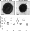
FIG. 1.
FIG. 1.
Capsular thickness of opaque- and transparent-phase variants of TIGR4(−)+ capsular serotype-switched strains. (A) Representative EM images of TIGR4(−)+6A opaque- and transparent-phase variants. (B) Medians and IQRs of capsule thickness (nm) measured by EM for opaque (clear boxes)- and transparent (diagonally shaded boxes)-phase variants of TIGR4(−)+6A, +7F, +23F, and +4 strains. *, Ρ < 0.01; **, Ρ < 0.001; ***, Ρ < 0.0001 (Mann-Whitney U test).
FIG. 2.
FIG. 2.
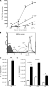
C3b/iC3b deposition on TIGR4(−)+ strains. (A) Mean FIs (measured in arbitrary units) of C3b/iC3b deposition measured using flow cytometry on TIGR4(−)+ strains expressing capsular serotypes 6A (▪), 7F (▴), 23F (▿), and 4 (○) in increasing concentrations of human serum. Error bars represent SDs and when not present are too small to be visible outside the symbol. **, Ρ < 0.001 (compared to the TIGR4(−)+4 strain; ANOVA with post-hoc tests). (B) Examples of flow cytometry histograms for C3b/iC3b deposition on TIGR4(−)+ capsular serotype-switched strains in 100% human serum. Gray shading indicates the results for bacteria incubated in PBS alone. (C and D) Mean FIs of C3b/iC3b deposition measured using flow cytometry on opaque- and transparent-phase variants of TIGR4(−)+ strains expressing capsular serotypes 6A (C) and on 7F, 23F, and 4 (D) in 25% human serum. Error bars represent SDs. *, Ρ < 0.01; **, Ρ < 0.001 (Student unpaired t tests).
FIG. 3.
FIG. 3.
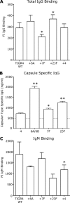
Ab binding to TIGR4(−)+ strains in human serum used for the C3b/iC3b and phagocytosis assays. (A) Mean FIs of total IgG binding to TIGR4(−)+6A, +7F, +23F, and +4 strains measured using flow cytometry. Treatment of the serum with IdeS prior to incubation with S. pneumoniae abolished IgG binding as detected by flow cytometry. The mean FI for total IgG binding to TIGR4 was 480 ± 40 in BSA-treated serum versus 30 ± 12 in IdeS-treated serum. (B) Mean amounts (ng/ml) of capsule-specific IgG measured using ELISA against capsular serotypes 4, 6A/6B (the assay is unable to distinguish between these subtypes), 7F, and 23F. (C) Mean FIs of total IgM binding to TIGR4(−)+6A, +7F, +23F, and +4 strains measured using flow cytometry. In all of the panels, the error bars represent SDs. *, Ρ < 0.01; **, Ρ < 0.001 [compared to the TIGR4(−)+4 strain; ANOVA with post-hoc tests]. WT, wild type.
FIG. 4.
FIG. 4.
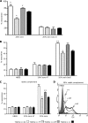
IgG-independent C3b/iC3b deposition on TIGR4(−)+ capsular serotype-switched strains. (A) Mean FIs of C3b/iC3b deposition measured using flow cytometry on the TIGR4(−)+ capsular serotype-switched strains in 50% human serum treated with IdeS to deplete IgG, with an example of the corresponding flow cytometry histograms. (B) Mean FIs of C3b/iC3b deposition measured using flow cytometry on the TIGR4(−)+ capsular serotype-switched strains in 25% baby rabbit complement, with an example of the corresponding flow cytometry histograms. In all of the panels, the error bars represent SDs. **, Ρ < 0.001 [compared to the TIGR4(−)+4 strain; ANOVA with post-hoc tests]. Gray shading represents flow cytometry histograms for bacteria incubated in PBS rather than serum.
FIG. 5.
FIG. 5.
Effect of the capsular serotype on human neutrophil phagocytosis measured using flow cytometry. (A) Mean (SD) proportions of neutrophils associated with fluorescent (labeled with FAMSE) TIGR4(−)+6A (white columns), +7F (stippled columns), +23F (gray columns), and +4 (black columns) strains after incubation in 20% human serum or 20% human serum treated with 5 μM cytochalasin D (Cyto D). (B) Mean (SD) proportions of neutrophils associated with fluorescent (labeled with FAMSE) TIGR4(−)+6A (white columns), +7F (stippled columns), +23F (gray columns), and +4 (black columns) strains after incubation in Hanks balanced salt solution (HBSS) buffer, heat-treated (HT) 20% human serum, or 20% human IgG-depleted serum (using treatment with IdeS). (C) Mean (SD) proportions of neutrophils associated with fluorescent TIGR4(−)+6A (white columns), +7F (stippled columns), +23F (gray columns), and +4 (black columns) strains after incubation in HBSS buffer, heat-treated 20% baby rabbit complement, or 20% baby rabbit complement. For panels A, B, and C, a single asterisk indicates a Ρ value of <0.01 and double asterisks indicate a Ρ value of <0.001 (compared to the TIGR4(−)+4 strain; ANOVAs with post-hoc tests). (D) Examples of flow cytometry histograms for the association of TIGR4(−)+4, +6A, and +23F capsular serotype-switched strains with neutrophils after opsonization with 20% baby rabbit complement. Gray shading indicates the results for bacteria incubated in HBSS alone. The histogram for the TIGR4(−)+7F strain is not included for ease of interpretation and because this strain had no significant differences from the TIGR4(−)+4 strain.
FIG. 6.
FIG. 6.
Virulence of capsular serotype-switched TIGR4(−)+ strains in mice. (A) Mean (SD) proportions of human neutrophils associated with fluorescent (labeled with FAMSE) TIGR4(−)+6A (white columns), +7F (stippled columns), +23F (gray columns), and +4 (black columns) strains after incubation in various concentrations of mouse serum and in heat-treated (HT) mouse serum. Error bars represent SDs. *, Ρ < 0.01; **, Ρ < 0.001 (compared to the TIGR4(−)+4 strain; ANOVAs with post-hoc tests). (B and C) Virulence in a mouse model of septicemia. Each symbol represents the median number of bacterial CFU recovered from an individual mouse spleen (B) or blood (C) 24 h after i.p. inoculation with 2,000 CFU of the TIGR4(−)+6A, +7F, +23F, or +4 strain. Bars represent median numbers of CFU of each bacterial strains, and very similar data were obtained in a repeat experiment. For both blood and spleen, a P value of 0.0005 (Kruskal-Wallis test) was obtained for the overall comparison between strains. P values for individual strains compared to the TIGR4(−)+4 strain are given above the symbols for the strains (Dunn's multiple-comparison test).

Similar articles

Cited by

References

    1. Aanensen, D. M., A. Mavroidi, S. D. Bentley, P. R. Reeves, and B. G. Spratt. 2007. Predicted functions and linkage specificities of the products of the Streptococcus pneumoniae capsular biosynthetic loci. J. Bacteriol. 189:7856-7876. - PMC - PubMed
    1. Abeyta, M., G. G. Hardy, and J. Yother. 2003. Genetic alteration of capsule type but not PspA type affects accessibility of surface-bound complement and surface antigens of Streptococcus pneumoniae. Infect. Immun. 71:218-225. - PMC - PubMed
    1. Arredouani, M., Z. Yang, Y. Ning, G. Qin, R. Soininen, K. Tryggvason, and L. Kobzik. 2004. The scavenger receptor MARCO is required for lung defense against pneumococcal pneumonia and inhaled particles. J. Exp. Med. 200:267-272. - PMC - PubMed
    1. Austrian, R. 1981. Some observations on the pneumococcus and on the current status of pneumococcal disease and its prevention. Rev. Infect. Dis. 3(Suppl.):S1-S17. - PubMed
    1. Austrian, R., H. P. Bernheimer, E. E. Smith, and G. T. Mills. 1959. Simultaneous production of two capsular polysaccharides by pneumococcus. II. The genetic and biochemical bases of binary capsulation. J. Exp. Med. 110:585-602. - PMC - PubMed

Publication types

LinkOut - more resources