. 2010 Mar;139(3):613-21.
doi: 10.1530/REP-09-0284. Epub 2009 Nov 30.

Aquaporin 9 expression in the developing rat epididymis is modulated by steroid hormones

Affiliations

Aquaporin 9 expression in the developing rat epididymis is modulated by steroid hormones

Núria M Pastor-Soler et al. Reproduction. 2010 Mar.

Abstract

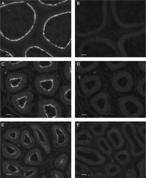
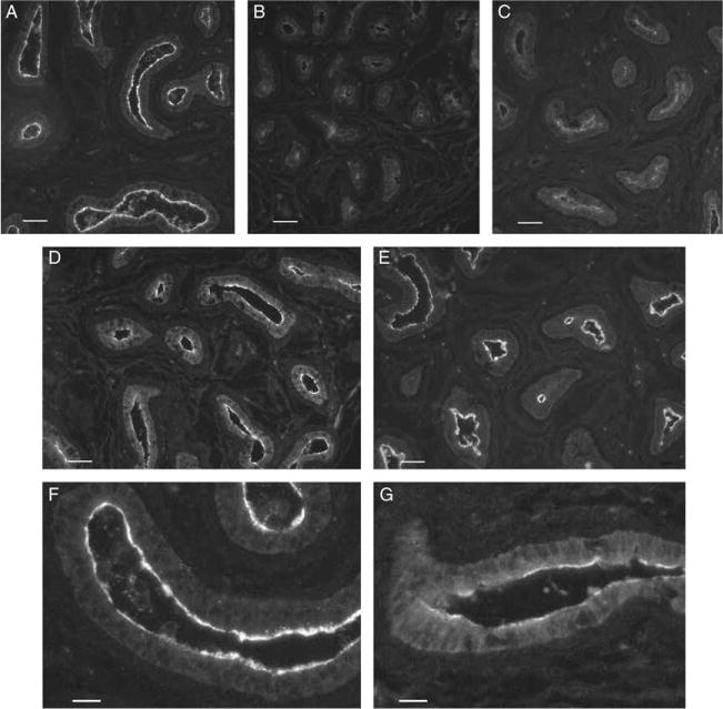
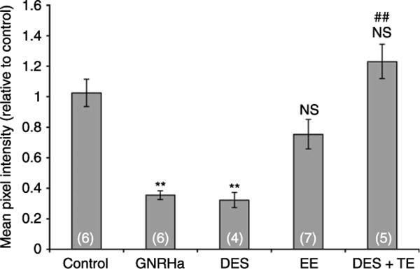
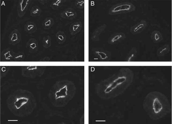
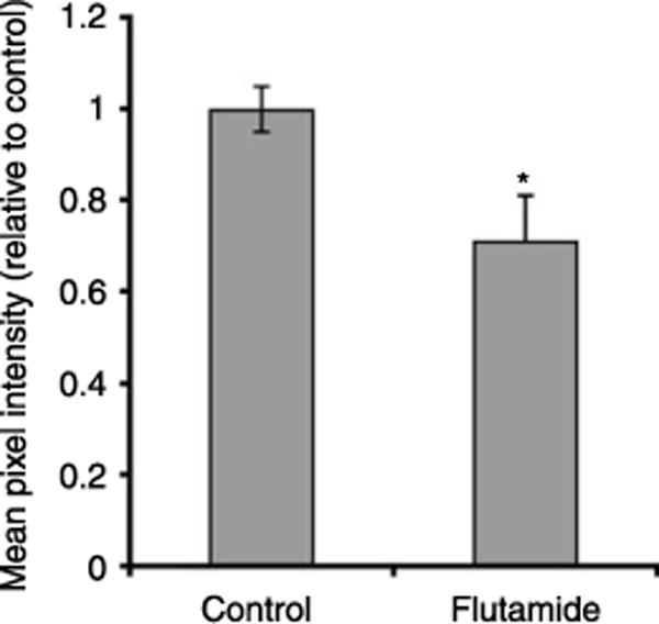
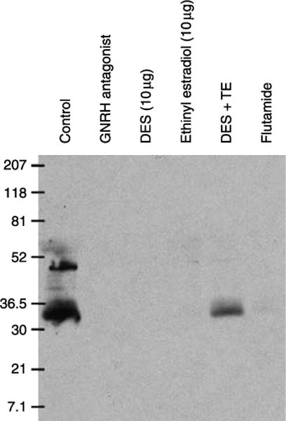
Fluid and solute transport across the epithelium of the male excurrent duct is important for sperm maturation and storage. Aquaporin 9 (AQP9), which allows permeation of water and neutral solutes, is abundant throughout the male reproductive tract, where it is expressed at the apical membrane of rat epididymal principal cells as early as at 1 week of age. We evaluated the effect of neonatal exposure to: 1) a GNRH antagonist (GNRHa); 2) diethylstilbestrol (DES); 3) ethinyl estradiol (EE); 4) DES plus testosterone (DES+TE); and 5) the anti-androgen flutamide on AQP9 expression in the epididymis of peripubertal rats. Control groups received the vehicle alone. In 25-day-old rats, quantification of the mean pixel intensity of immunofluorescence-stained sections showed a significant decrease in AQP9 staining in the apical membrane of epididymal principal cells after treatments with GNRHa, DES, or flutamide, compared to controls. These results were confirmed by western blotting. While EE induced a marked decrease in AQP9 levels by western blotting, the decrease in AQP9-associated fluorescence was not significant compared to controls. DES+TE-treated rats showed levels of AQP9 protein similar to controls, indicating maintenance of AQP9 expression by testosterone treatment in the presence of DES. Our data show that expression of AQP9 in the developing rat epididymis is downregulated by neonatal DES, GNRHa, EE, and flutamide, and that the effects mediated by estrogens can be prevented by testosterone administration.


Conflict of interest statement

Declaration of interest

The authors declare that there is no conflict of interest that could be perceived as prejudicing the impartiality of the research reported.

Figures

Figure 1
Figure 1
AQP9 immunostaining in the proximal cauda epididymidis of an adult rat (A), a 4-week-old rat (C), and a 3-week-old rat (E). A bright staining was detected in the apical membrane of principal cells at all time points. The staining reached maximal intensity, comparable to adult levels, between the third and fourth postnatal weeks. (B, D and F) Pre-incubation of the antibody with the AQP9 peptide completely abolished the staining at all time points. B, control; D, 4-week-old; F, 3-week-old epididymis. Bars=25 μm.
Figure 2
Figure 2
AQP9 immunostaining in the cauda epididymidis of 25-day-old rats after neonatal treatment with (A) vehicle (control); (B) GNRHa; (C) DES; (D) EE; and (E) DES+TE. (F and G) Higher magnification pictures of control and EE-treated animals respectively. GNRHa and DES treatments caused a marked decrease in apical AQP9 staining. EE-treated animals showed a moderate decrease in AQP9 immunofluorescence labeling. Administration of TE maintained AQP9 expression in DES-treated rats at control levels. Digital images were acquired using identical parameters. (A–E) Bars=40 μm. (F and G) Bars=15 μm.
Figure 3
Figure 3
Quantification of AQP9-associated fluorescence staining in the cauda epididymidis of 25-day-old control and treated rats (groups represented in Fig. 1). Significant decreases in AQP9 staining intensity were induced by GNRHa and DES. EE also induced a reduction in AQP9 staining but the difference was not significant versus control. Co-administration of testosterone with DES completely restored AQP9 staining compared to DES alone, with AQP9 staining intensity comparable to control. Data are expressed relative to control, and represent the means±S.E.M. obtained from 4 to 7 animals per group. **P<0.0005 versus control; ##P<0.005 versus DES; NS, not significant versus control.
Figure 4
Figure 4
Downregulation of AQP9 expression in the cauda epididymidis of 25-day-old rats by the anti-androgen flutamide. Immunofluorescence for AQP9 in the cauda epididymidis from a control rat (A) and a flutamide-treated rat (B) showing a marked decrease in apical AQP9 staining in the flutamide-treated animal. Panels C and D are higher magnification representations of some of the tubules shown in A and B respectively. Digital images were acquired using identical parameters. Bars=25 μm.
Figure 5
Figure 5
Quantification of AQP9-associated fluorescence staining in the cauda epididymidis of control and flutamide-treated 25-day-old rats. The mean pixel intensity of AQP9-associated fluorescence in the apical pole of principal cells was markedly decreased following flutamide treatment. Data are expressed relative to control and represent the means±S.E.M. obtained from three animals per group. *P<0.05 versus control.
Figure 6
Figure 6
Western blot showing downregulation of AQP9 in epididymis of 25-day-old rats by different hormonal treatments. A strong band at ~30–35 kDa is present in total epididymal homogenate membrane of control animals. Additional, higher molecular weight bands probably represent different glycosylation states of AQP9. AQP9 was not detectable in the GNRHa-, DES- and EE-treated groups. In the DES+TE-treated animals, a brighter AQP9 band was detected compared to DES treatment alone. A very faint AQP9 band was detected in the flutamide-treated group. Each well was loaded with the same amount of protein (Fisher et al. 2002).

Similar articles

Cited by

References

    1. Atanassova N, McKinnell C, Walker M, Turner KJ, Fisher JS, Morley M, Millar MR, Groome NP, Sharpe RM. Permanent effects of neonatal estrogen exposure in rats on reproductive hormone levels, Sertoli cell number, and the efficiency of spermatogenesis in adulthood. Endocrinology. 1999;140:5364–5373. - PubMed
    1. Atanassova N, McKinnell C, Fisher J, Sharpe RM. Neonatal treatment of rats with diethylstilboestrol (DES) induces stromal–epithelial abnormalities of the vas deferens and cauda epididymis in adulthood following delayed basal cell development. Reproduction. 2005;129:589–601. - PubMed
    1. Badran HH, Hermo LS. Expression and regulation of aquaporins 1, 8, and 9 in the testis, efferent ducts, and epididymis of adult rats and during postnatal development. Journal of Andrology. 2002;23:358–373. - PubMed
    1. Bagnis C, Marsolais M, Biemesderfer D, Laprade R, Breton S. Na+/H+-exchange activity and immunolocalization of NHE3 in rat epididymis. American Journal of Physiology Renal Physiology. 2001;280:F426–F436. - PubMed
    1. Belleannee C, Da Silva N, Shum WW, Marsolais M, Laprade R, Brown D, Breton S. Segmental expression of the bradykinin type 2 receptor in rat efferent ducts and epididymis and its role in the regulation of aquaporin 9. Biology of Reproduction. 2009;80:134–143. - PMC - PubMed

Publication types

MeSH terms

LinkOut - more resources